TMJ diagnosis starts with your story and a hands-on exam, not with a “scan just in case.” I am Dr. Maksut Behruzoglu, a specialist orthodontist in Dubai; in TMJ diagnosis and TMJ treatment visits I collect the timeline of pain, clicking, limited opening, morning muscle fatigue, clenching habits, and sleep. Then I look at the face, measure jaw motion, palpate joints and chewing muscles, and check the bite and contacts. Imaging — panoramic films, periapicals, CBCT, MRI, sometimes a bite record and a scan — I order for a specific question, not as a default bundle. Recent guidance on temporomandibular disorders agrees: the clinical picture leads, imaging refines. I do not diagnose a click from a photo. Below — what I check in the chair, when each study earns its place, and how we separate a joint story from tooth pain, sinus issues, or another kind of headache.
Why diagnosis beats “treat the click”
Friends hear a click at breakfast. A search thread labels it “TMJ” before anyone has measured opening. I hear that label in English, Russian, and Turkish in the same week. A painless click with free opening is one portrait. Pain in front of the ear, morning lock feeling, joint locking, temple pain with tired masseters is another. Glue them under one word and jump to a splint or braces and you buy disappointment.
Diagnosis answers three working questions. First: where do the symptoms live — joint, muscles, teeth, nearby face and neck structures, or a mix. Second: are there red flags that need another specialty now. Third: which tools fit today — watchful waiting, a splint, physiotherapy, orthodontics, or shared work with an oral surgeon or neurologist. Without those answers an appliance is décor.
Dubai adds a transfer layer: films from several countries, fragmented reports, “they already said the joint is destroyed.” I respect prior documents and still build my own clinical base. Second opinions are welcome. AED figures come after a plan with a price-list date if cost enters the talk. Another trap is treating one symptom in isolation. You ask to remove a click and omit night grinding, one-sided chewing, a recent high crown, or unfinished aligner retention. I pull the timeline back one to two years. Without the timeline it is easy to treat someone else’s hypothesis. Below — how the history becomes an exam.
History first: what I ask before I touch the face
Patients often arrive with one complaint: a click, temple pain, “my jaw sticks,” wear, “my dentist said the bite is guilty.” I widen the picture: time of day, food triggers, stress, sleep, trauma, past orthodontics, chewing side preference, night grinding reported by a partner. Answers build hypotheses before hands meet the face.
Pain that builds toward evening after calls and a clenched jaw on video leans muscular. Pain with morning limitation that “works itself open” over an hour can point to inflammation or internal disc dysfunction. Sharp tooth pain to cold points to a restorative colleague even if you booked a “joint visit.” Sinus disease, migraine, neuralgia are separate roads; I do not claim them for orthodontics out of convenience.
Sleep and stress enter the history without lectures. I do not preach “just relax.” I record facts: night grinding, sleep apnea notes from a physician, jet lag, deadline seasons. Those loads change muscle and joint demand. They do not cancel a bite exam and they do not make braces an automatic headache cure.
Medications, new crowns, a recent wisdom-tooth removal, a sports hit to the jaw can shift the picture. Expats often move care between clinics and lose files. Bring what you have; I mark the gaps. Laptop and phone posture matter too: head forward, shoulders up, jaw clenched for hours. That is not a TMJ diagnosis by itself; it is load on temporal and neck muscles. Temple pain after online meetings with little chewing pain raises the muscle contribution. Pain on a wide yawn and hard food keeps the joint contribution on the table. Below — how complaints become a physical exam.
Clinical exam of the face, joint, and muscles
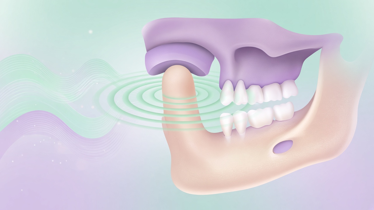
The exam is the core of TMJ diagnosis. I look at facial symmetry at rest and in smile, the midline, lip posture, and signs of high tone in masseter and temporalis. Then I ask you to open slowly to a comfortable limit while we watch pain. I measure opening, note deviation, and record sounds: click, crepitus, “sand.” I palpate the joint in front of the ear and, when useful, via the external auditory canal. Muscles — temporalis, masseter, pterygoids as accessible — with matched force left to right. Bite and contacts enter the same visit without the automatic formula “crooked teeth equal joint disease.” Neck and shoulders join the talk if pain refers there: I do not diagnose the cervical spine, but I note the link and refer. Below — three exam layers patients often mix up.
Range, deviation, and locking
Adult opening ranges sit in a clinic’s familiar band; dynamics and pain matter more than one textbook number. Deviation on opening can travel with disc dysfunction or muscle imbalance. Locking means opening or closing stops short and you feel stuck. An acute lock needs calm priority: not always “just stress,” not always urgent surgery. I test whether soft chairside manoeuvres free motion, whether imaging is needed, and whom to involve.
Patients mid-orthodontics sometimes blame wires or elastics. Sometimes timing matches; sometimes it coincides. I rebuild chronology against activations, trauma, and bite-height changes after restorations. Without chronology it is easy to blame the appliance.
Side and forward movements belong in the exam. Limited laterotrusion, pain on protrusion, asymmetric travel add to the opening story. I ask which chewing side feels easy and which feels wrong: a “favourite” side can be a bypass of a joint or a tooth. Face and arch photos fix a baseline so the next visit compares a frame, not memory.
Joint sounds: click and crepitus
A click often links to disc displacement with reduction: the disc shifts, then seats with a sound. Crepitus — a coarser grinding noise — can signal degenerative surface change. Sound alone is not a “destroyed joint” verdict and not a surgical ticket. I link sound to pain, limitation, and imaging when indicated. Painless clicks are watched for years without aggression; painful progressive clicks take another path.
Patients ask braces to “switch off” the click. Orthodontics changes tooth position and contacts; it is not a universal sound switch. I say that before any promise in TMJ treatment.
Muscle map and referred pain
Temporalis often refers to the temple and feels like a “bite headache.” Masseter refers to the cheek and jaw angle. Pterygoids join lateral motion and opening; their trouble mimics ear or tooth pain. I note whether muscle palpation reproduces the familiar pain more than joint palpation. That ranking steers the first step: unloading and a splint, sleep and clenching hygiene, physiotherapy — or deeper joint imaging.
Referred molar pain without decay is a classic trap. A dentist drills a healthy tooth, pain stays, trust collapses. Bring tooth films; we discuss why a muscle source may lead if the clinic points there. The tooth decision still belongs to the restorative clinician on site.
Tender points and referral into temple or teeth help separate a muscle map from a pure joint map. A crowded arch can sit beside a quiet joint; perfectly straight teeth do not guarantee quiet muscles. I say what I see and where the hypothesis is still thin.
Bite and occlusion inside a TMJ work-up
The bite is part of TMJ diagnosis, not the sole culprit. I check jaw relationship in habitual occlusion, crossbites, deep bite with palate trauma, open bite with missing chewing contacts, lateral shifts. Articulating paper or a digital scan shows where a tooth meets early. An early contact raises muscle activity in some people; in others the same contact lives for years without pain. Wear facets, chips, cracks mark overload, not automatic proof that “the bite caused the headache.” Bite height after worn teeth or high crowns I judge carefully: relief after contact refinement happens; a universal temple cure does not. I test hypotheses and record tissue response rather than selling a pre-chosen appliance. I look for bite–joint links across timeline, muscles, and contacts together, not from one pretty scan. Below — three practical layers before force.
Habitual occlusion and a forced position
Habitual occlusion is how you close every day. A forced position is the jaw sliding sideways or forward to find a comfortable contact around an interference. I ask for your usual close, then guide toward a more neutral contact when it is safe and informative. The gap between habit and a more central seat explains why some people feel a “jump” and why films can surprise them.
Adults with years of one-sided chewing often show muscle and wear asymmetry. That is not a verdict of internal disc derangement. It is a working hypothesis for further tests.
Adults with long orthodontic history ask: “my old bite drifted after the retainer — did that wreck the joint?” Drifted teeth change contacts; joints answer load in different ways. A straight line from relapse to joint destruction is usually too crude. I read current symptoms, muscles, range, and imaging when indicated before I tie dental instability to a joint complaint.
Contacts, paper, and digital scans
Articulating paper remains a fast clinical tool. A digital scan gives a model for discussion and for splint work. I show thicker contact marks and link them to complaints when the link is plausible. If muscles are quiet and contacts look “imperfect,” I do not treat a contact map for cosmetic neatness.
In Dubai, patients bring splints from other countries. I judge fit, height, and wear marks, not the plastic brand. A poor splint becomes a pain source; a perfect-looking splint without a diagnosis is also a risk.
Orthodontics can redistribute contacts; it does not treat night grinding as a sleep-medicine or stress topic by itself. Overload may come from clenching, stress, apnea, or a mix. I record overload markers and weigh each layer instead of hanging one label.
When orthodontics is early in the talk
Braces or aligners change occlusion over months. With acute joint pain or a strong muscle syndrome I often pause force or delay start until the acute phase settles. That is sequence, not a lifelong ban. Patients who came to “straighten teeth and cure the joint in one go” hear a clear fork: orthodontics solves dental and skeletal goals when indicated; joints and muscles need their own work-up and sometimes a separate course.
Which studies I order and when
Studies confirm or refine a clinical hypothesis. I do not MRI every click and I do not refuse imaging when red flags sit on the table. A panoramic film (OPG) surveys teeth, jawbones, and a coarse view of condyles. Periapicals close tooth pain. CBCT shows bony joint contours in three dimensions when indicated. MRI sees disc, soft tissue, and effusion better. Method choice answers a question; it is not a clinic status symbol. Dose, cost, Dubai access, and quality of old foreign films enter the decision. AAOMR and dental imaging guides stress that CBCT is justified when a two-dimensional method fails the question; for the disc, MRI remains the leading soft-tissue view. I explain the difference before referral. I write the referral with a clinical question. Below — three modalities in plain language.
OPG and periapical films
I often use a panoramic film as a survey: impacted teeth, coarse condyle asymmetry, bone around teeth, large findings. It does not replace MRI for a disc question. Periapicals and vitality tests close odontogenic pain. A patient with “joint” pain and deep molar decay goes to restorative care first — sequence, not dismissal.
Old OPGs from another country help for change over time if quality is readable. A blurred five-year-old film is not today’s joint status.
CBCT: when three-dimensional bone is needed
I order CBCT when the question is bony joint contours, trauma, marked asymmetry, surgical planning, or when a 2D film is not enough. I do not CBCT for archive beauty. Dose is higher than a periapical; benefit must clear the bar. The referral states what we seek, which joint, and a protocol the radiologist can match when possible.
Patients confuse facial CBCT with MRI. I draw the line: CBCT — bone; MRI — soft tissue and disc. Both may be needed by different people at different times.
MRI of the joint: disc and soft tissues
MRI of the temporomandibular joint is the method of choice when we need disc position, effusion signs, and soft-tissue status in pain, locking, or suspected internal derangement. I refer after the exam, with a written question. I read the radiology report beside your symptoms: disc displacement on film without pain or limitation is not an automatic surgery sentence.
Dubai centres vary. I prefer teams that run dedicated TMJ protocols, not a generic head scan missing the needed slices. If you bring a prior MRI disc, bring the protocol description too.
Imaging map in one table
The table below is the working map I keep in mind at consultation. It is not a price list and not a mandatory package. Your set comes only after examination; timelines and fees depend on the imaging centre and the price-list date.
| Clinical question | First step | When we go deeper | What we usually seek | What the method does not do |
|---|---|---|---|---|
| Tooth pain vs joint | Exam + periapical / vitality tests | OPG for a wider view | Decay, pulpitis, crack | Does not show the TMJ disc |
| Jaw survey, coarse joint | OPG | Condyle asymmetry, trauma | Teeth, bone, condyle shape | Weak on disc and soft tissue |
| Joint bone in 3D | CBCT when indicated | After clinic, not “for everyone” | Contours, erosions, osteophytes | Does not replace MRI of the disc |
| Disc, effusion, soft tissue | MRI when indicated | Pain, lock, suspected derangement | Disc position, fluid | Does not treat; only a picture |
| Contacts and splint | Scan / impression + clinic | When unloading is planned | Occlusion model | Not a TMJ diagnosis by itself |
| Muscle pain, no red flags | Clinic ± splint trial | Imaging if no response | Triggers, symmetry | A scan is not required at once |
| Temple headache | History + muscles + bite | Neurologist for migraine pattern | Link to chewing / clenching | Orthodontist does not replace neurology |
Patients often ask to “do everything now.” A package without a question adds dose, cost, and anxiety about findings that do not explain the complaint. I order the next film when the answer will change the plan: splint or not, force now or later, referral to surgery or neurology. If the clinic is muscular and red flags are absent, we start with exam and unloading, then add imaging if there is no response or new signs appear.
Differential diagnosis: what masquerades as TMJ
Not every pain in front of the ear is the joint. Otitis, ear-canal problems, sinus disease, neuralgia, salivary pathology, tumours (rare, but red flags matter), cervical sources — all cross the zone you point to. I am an orthodontist; when doubt rises I refer to ENT, neurology, oral and maxillofacial surgery, or a GP. That is a competence border, not a brush-off.
Referred tooth pain and muscle pain into a tooth confuse even strong teams. Repeated root canals with no relief are a reason to return to the muscle and joint map. The reverse — long “joint massage” during acute pulpitis — wastes time and risks.
Tension-type headache and migraine have their own criteria. If temples pulse with light sensitivity, nausea, and aura, the neurology road leads even if the bite is imperfect. I can help unload muscles when indicated; I do not replace a neurologist with a promise that “straight teeth end migraine.”
Pain from all-day gum chewing at work differs from pain on one cinema yawn. The first often leans muscle and habit; the second leans joint and range. Both can coexist. TMJ diagnosis exists so we do not collapse everything into one search label.
Red flags: progressive facial asymmetry, neurologic deficit, unexplained weight loss, night pain with systemic signs, trauma with fracture risk — urgent routing, not an aesthetic tray talk. These cases are rarer than classic muscle syndromes and still must not be missed. If a red flag appears in the orthodontic chair, I pause cosmetic planning and move you to the right contour the same day when Dubai logistics allow.
After the diagnosis: a plan without guarantees
When hypotheses are gathered I name a working diagnosis in plain words: muscle component, joint component, mixed, odontogenic contribution, habit contribution. Then steps: observation and clenching hygiene; splint therapy when indicated; physiotherapy; coordination with other specialists; orthodontics if a dental goal is real and the acute phase is controlled. Timelines and response are individual. I do not guarantee “the click vanishes in a month.”
I document baseline ranges, photos, films when indicated, and complaints in your words. At review we compare to your start, not to an advert ideal. If the plan does not shift the picture, we change the hypothesis — ordinary medicine, not a brand failure.
In TMJ treatment, orthodontics is one tool beside others, not the only one. Patients who expect braces to cure every headache hear a calm refusal of that promise already at diagnosis. An honest border keeps trust across years of care.
A splint, when indicated, is diagnostic and therapeutic at once: it unloads, protects teeth from grinding, and sometimes clarifies whether the system answers to contact change. If weeks of wear do not move the picture, we do not raise height at random; we revise the hypothesis. If it moves, we record what worked and decide whether orthodontics, restorations, or support alone come next.
Children and teens with clicks need separate caution: growth, past sucking habits, ENT issues, appliances. I do not copy the adult protocol one-to-one. Parents hear that “it clicks like the adult video” is not a child’s diagnosis. Exam, follow-up, sometimes observation without an appliance — a normal path.
Patients after orthognathic surgery bring another level: the joint has lived through osteotomies, intermaxillary fixation, elastics. I build diagnosis with the surgeon who operated or who follows now. Solo “bite upgrades” with trays without the team are risky in those cases.
UAE insurance covers TMJ imaging unevenly. I state medical need; cover confirmation comes from the policy administrator or third-party administrator (TPA). If you self-pay, we rank priority: which film changes the plan now, which can wait. Honest sequence saves money and anxiety.
Repeat diagnosis months later is needed when symptoms change, an appliance fails to answer, or a new red flag appears. I do not repeat MRI “for the chart” every three months without clinic. Symptom, range, and muscle-map change often inform more than an extra frame.
Patients ask whether one visit can “close the topic.” Sometimes yes: the muscle pattern is clear, red flags are absent, an unloading plan is agreed. Sometimes no: films, a neurology note, a splint try-in, a two-to-four-week review. I name the next step aloud so expectation matches medicine, not a “diagnosis in fifteen minutes” advert.
FAQ on TMJ diagnosis
Does everyone with a clicking joint need an MRI?
No. A painless click without limited opening is often watched clinically. I order MRI when the clinic asks about the disc, locking, effusion, or when soft-tissue detail will change the plan. A scan without a question adds anxiety and cost.
Can an OPG alone diagnose TMJ disorders?
An OPG gives a survey and a coarse look at bony contours, but it sees disc and soft tissue poorly. Diagnosis rests on history and exam; imaging refines. One panoramic frame rarely finishes a joint conclusion.
CBCT or MRI first?
Clinic first. If the question is bone, CBCT when indicated. If the question is disc and soft tissue, MRI. Sometimes both at different times. I do not pick the “more modern” method for status; I pick the answer to your question.
Is TMJ diagnosis the same as an orthodontic plan?
No. An orthodontic plan moves teeth toward bite and aesthetic goals. TMJ diagnosis asks whether force is safe to start now and whether a separate joint phase is needed. Courses may run in sequence or in parallel with limits. Decision after examination.
How long is a diagnostic visit?
Usually a full consultation with history, exam, measurements, and discussion. If films are taken elsewhere, a second step follows once files arrive. Exact time depends on complaint complexity and the documents you bring.
What should I bring to a Dubai visit?
A timed symptom list, prior films on disc or cloud, surgeon and neurology notes, splints if you have them, medication list. Translation helps, but raw files beat nothing. I will mark gaps and say what is still missing.
Can a GP dentist or ENT replace the orthodontist here?
Each closes a contour. The dentist rules out the tooth; ENT covers ear and sinuses; neurology covers primary headaches; the orthodontist covers bite, chewing muscles, and links to an orthodontic plan. Mixed pictures need a team. One specialty rarely covers every mask.
Do you diagnose online from video?
No. Video helps gather history and urgency, but palpation, range, contacts, and imaging decisions need an in-person exam. Online I can suggest whom to see sooner; a working diagnosis happens in the chair.