A 3D bite model shows your teeth as they are today and the position I propose to move them toward. I am Dr. Maksut Behruzoglu, a specialist orthodontist in Dubai, and I use the model at consultation to explain the diagnosis, compare treatment options, and agree on a goal. The final frame of the model shows the orthodontist's intention. It cannot show how your bone, gums, and roots will respond, how many hours a day you'll wear your aligners, or how a year of business trips, holidays, and colds will go. Accuracy studies of clear aligners show that teeth reach, on average, about half of the planned movement for some movement types, and several studies report that most aligner patients need at least one refinement. That is why in 3D bite modeling I name the difficult teeth up front and show where the plan leaves room for corrections. This article covers how I work with the model, why simulation and reality diverge, which part of the result depends on you, and how to read the final frame without inflated expectations.
What a 3D bite model is and how it's built
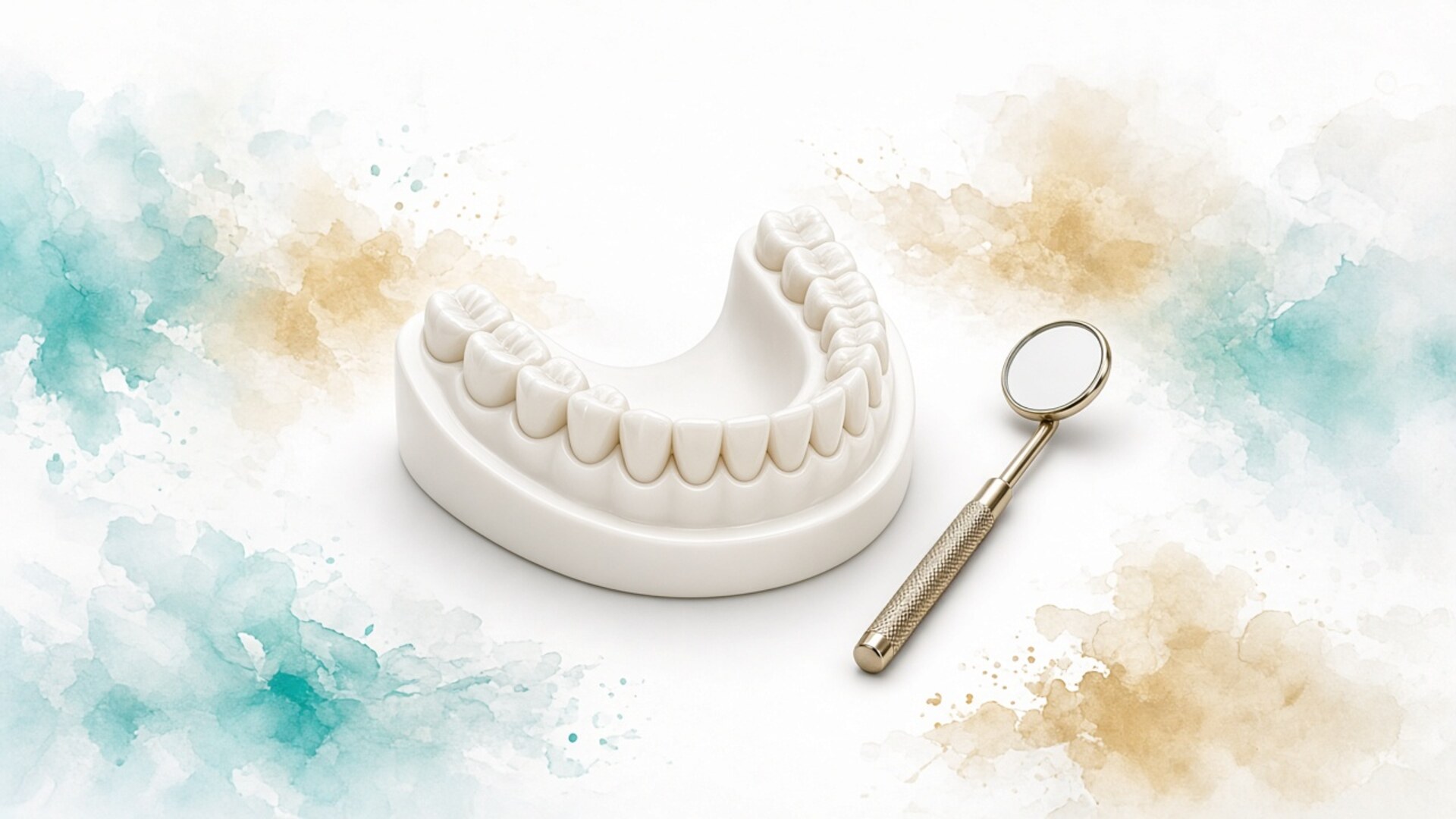
A 3D bite model is a digital copy of your teeth and gums built from an intraoral scan. The scanner captures the surfaces of the upper and lower arches and records how your teeth meet, and the software builds a model you can rotate, zoom, and section. This used to require impressions and plaster casts. Today a scan takes a few minutes and involves no tray of paste in your mouth.
The starting model is a record. It captures the position of every tooth, the arch form, gaps, crowding, and how the jaws relate at the recorded bite. It can be compared with a scan taken six months later to see what changed. For patients who relocate or switch orthodontists, it is a convenient way to hand over the treatment history.
From the starting model I build the planned one. The software splits the model into individual teeth, and I move them toward the target position and set the route: how many steps, in what order, with which auxiliaries. For clear aligners, each step becomes a separate tray. For braces, the model helps calculate space and bracket positions.
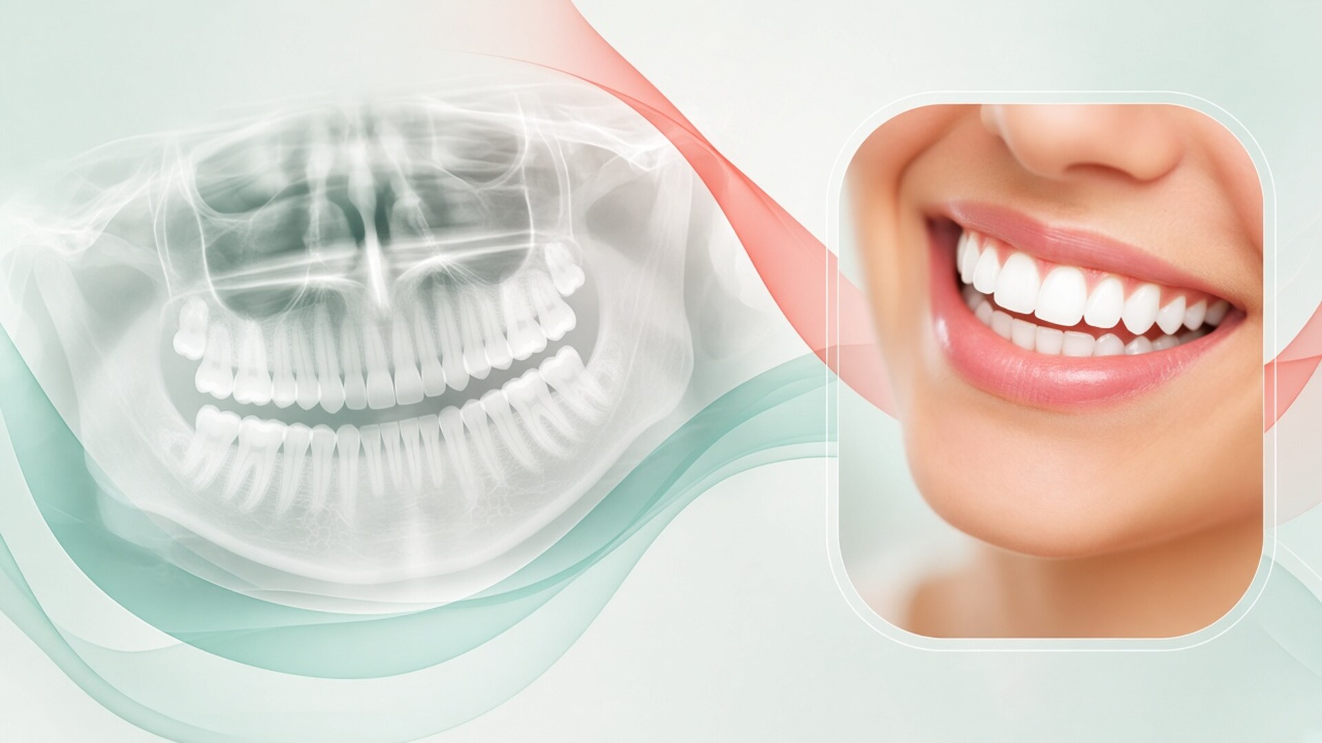
I add records the scan cannot capture. A panoramic X-ray shows roots and bone. A lateral cephalogram shows the jaw relationship and incisor angulation. Photos show your face and smile. When there is a clear indication, a CBCT scan gives a 3D view of roots and bone. Without these layers the model looks great yet stays incomplete: you see crowns and gums, while the structures holding each tooth stay out of frame.
Patients sometimes call the model "the computer's treatment plan," as if the software decides where the teeth go. The orthodontist decides. The software follows commands faithfully: if I place a tooth where it has no bone, the model will still show a pretty picture. So looking at the model without an orthodontist who explains the decisions tells you little. It looks equally convincing for a sound plan and a risky one.
The model itself has accuracy limits. Current intraoral scanners match conventional impressions for full-arch accuracy, but saliva, bleeding gums, and hard-to-reach back molars introduce error. The bite record captures one jaw position, the habitual one, which may differ from the position where the joints sit comfortably. I check the scan immediately and rescan any area that looks doubtful. An error in the starting model carries into every step after it, so five extra minutes in the chair can save months of corrections.
How I use the model during your consultation
The model helps me and my patients speak the same language. Words struggle to explain that the lower incisors are tipped forward a few degrees beyond normal and the back teeth meet half a cusp out of line. On screen you see it in a second. I rotate the model, show the bite from the side, from above, from the tongue side, and you see your teeth from the angle an orthodontist sees them. I use the model for three jobs: recording the starting point, comparing treatment options, and coordinating with other dentists and specialists. In each case the model supports a decision, and the decision itself comes out of our conversation. We put the goal into words first and only then translate it into tooth movements on screen.
Showing where you start
The first thing I do with the model is show what's there now: crowding in millimeters, tooth angulation, gaps, overbite depth, midline shift, arch asymmetry. You see the whole problem, not only the tooth that bothers you in the mirror.
Sometimes the patient's concern and the main bite problem differ. Someone comes in because of a crooked lateral incisor, and the model shows the lower jaw closing off-center with the back teeth overloaded. I explain what happens if we straighten that one incisor and leave the bite alone, and what full treatment would achieve. The choice stays with the patient, and it's made with eyes open.
I keep the starting model on file. A year into treatment people forget where they began, and comparing with that starting point shows real progress better than any description.
Comparing treatment options
Many cases can be treated in more than one way. Crowding can be resolved with enamel reduction, arch expansion, incisor tipping, or premolar extraction. A deep bite can be opened by intruding the front teeth or extruding the back teeth. On the model I build two or three versions and show them side by side.
Each version comes with my commentary: what it achieves, what it requires, what risks it carries. A non-extraction version can look just as good on screen as an extraction version while demanding incisor tipping the bone can't support. An option with elastics may finish sooner if you're ready to wear them. The model shows geometry; I add biology and lifestyle.
When two options are medically sound, we choose together. If one carries a clear risk to gums or roots, I say so and don't present it as an equal.
Working with your other dentists
The model is useful beyond the patient conversation. If neighboring teeth need uprighting before an implant, I send the model to the surgeon and the restorative dentist, and we agree on the space and the implant axis. If you plan veneers after orthodontics, the cosmetic dentist sees where the teeth will end up and plans veneer thickness.
For skeletal problems that need orthognathic surgery, the model helps the surgeon and orthodontist agree on pre-surgical tooth positions and the post-surgical bite. A periodontist can use the model and X-rays to judge where the gum is at risk and whether a graft is needed before or after tooth movement.
In Dubai many patients see several specialists at different practices. A shared model spares everyone the phone relay and lowers the risk that each dentist plans their piece in isolation.
Why a simulation isn't the result: biology
The model moves teeth; in real life, bone moves them. When an aligner or wire presses on a tooth, bone resorbs on one side of the root and builds up on the other. Cells run this process, and they work at their own pace. Age, bone density, root shape and length, gum health, certain medications, and general health all affect speed. Two patients with identical models and identical plans will see their teeth move differently. Within one mouth, incisors with slender roots move faster than molars with bulky roots. The planning software knows none of this. It divides the route into equal steps and shows smooth motion. I build in corrections I know from experience and research, yet I learn how your tissues actually respond only during treatment. Three biological factors drive the gap more than the rest.
How fast bone remodels
For aligners, a typical step per tray is about a quarter of a millimeter of linear movement and one to two degrees of rotation. Trays change every one to two weeks depending on the system and the plan. These numbers are set so that bone can keep up in most people.
Most people is not everyone. Adults have denser bone than teenagers, and it remodels more slowly. Conditions that affect bone metabolism and certain medications can slow things further. When a tooth can't keep up with the tray, the aligner stops seating fully and a gap appears between plastic and tooth. That is an early sign of lag.
I check aligner fit at every visit and ask patients to send photos if a tray sits loosely. Sometimes wearing a tray an extra week is enough. Sometimes we need a new scan and a new plan.
Roots, gums, and periodontal health
A root is anchored in bone by a ligament. As a tooth moves, the ligament and bone remodel, and the root tip sometimes shortens slightly, which is called root resorption. Mild resorption is common after orthodontic treatment and usually doesn't affect how the tooth functions. Severe resorption is less common, and the risk rises with long treatment, large root movements, previous trauma, and certain root shapes.
Gums respond in their own way. With a thin gum type and thin bone, tipping a tooth outward can lead to recession. The model shows the gum as a smooth pink surface and makes no prediction about its behavior. For patients with a history of periodontitis, I start orthodontics only after a periodontist has stabilized the gums, and I plan lighter forces.
None of this shows on the model. I monitor it with X-rays when indicated and with a gum check at every visit.
Growth and age
In children and teenagers the jaws are still growing, and growth direction can be predicted only roughly. The lower jaw may grow forward more than expected, and a bite that looked ideal on the model can change within two years. The model ignores growth: it moves teeth within jaws that stay still.
Adults don't grow, but they age. Over the years teeth drift even without orthodontics, especially the lower incisors. Gums and bone thin out. Restorations, crowns, and implants limit neighboring movement: an implant doesn't move at all, and the model has to account for that.
So with teenagers I revisit the plan as they grow, and with adults I set more modest goals where tissues are thin.
Your part of the work: wear time, elastics, hygiene
The model is designed for an ideal patient. The ideal patient wears aligners 22 hours a day, puts elastics back after every meal, never loses a tray on a flight, and brushes after every snack. Real people work, travel, get sick, get tired, and forget. I don't expect perfection, but I ask patients to see a simple link: every hour without aligners and every evening without elastics widens the gap between model and reality. After two months of patchy wear, trays seat with gaps, elastics haven't done their share, and the plan needs revising. With braces, less depends on the patient, yet elastics, hygiene, and intact brackets still rest on you. In aligner treatment planning I discuss your routine in advance and adapt the plan to real life. Three areas of patient responsibility affect the outcome most.
Wear time
An aligner works only while it's on your teeth. Most systems recommend 20 to 22 hours a day. That means trays come out only for meals, drinks other than water, and brushing. A latte at your desk stretched over an hour costs an hour of wear.
If you wear aligners 16 hours instead of 22, each tray does less work. The tooth doesn't reach its position, the next tray goes on tight, then loose. After a few trays the lag becomes visible. Some systems include wear indicators, but more often the orthodontist spots the problem from tray fit and photos.
I ask patients to be honest about how much they wear. If your schedule doesn't allow 22 hours, I can slow the tray changes or we can discuss braces.
Elastics and attachments
Inter-arch elastics correct how the jaws relate: they move back teeth forward or backward, close an open bite, and adjust the midline. The rule matches aligners: wear them nearly all the time. Elastics worn only at night deliver a small fraction of the force needed.
Attachments, small composite bumps on the teeth, give the aligner a grip so it can deliver force. If an attachment chips or wears down, the tray loses grip and the tooth stops moving as planned. Patients often don't notice a lost attachment. I ask you to check your teeth in the mirror once a week and let me know if one is missing.
With braces, brackets and ties play that role. A loose bracket switches the tooth out of the system until it's repaired.
Missed days, breakages, and travel
Dubai patients travel a lot: business trips, holidays, summers abroad. I send you off with the next tray and suggest keeping the previous one as a spare. If a tray breaks or gets lost, going back to the previous tray beats going without for several days.
Missed check-ups widen the gap too. At a visit I catch lag early, while it can be fixed without a new series. If you come in after four months instead of two, the lag has already piled up.
Illness, pregnancy, surgery, and stress can disrupt your routine. I don't lecture; I adjust the plan. We share the goal, and the route can change.
Refinements: why extra aligner rounds are normal
Refinements, additional aligner series based on a new scan, are a routine part of clear aligner treatment. When the first series ends, or when lag becomes noticeable, I take a new scan. The software builds a model from the current tooth positions, and I plan a short series to finish the remaining movements.
Refinements happen for several reasons. Some movements, such as rotating round teeth or extruding incisors, fall short of the plan. Teeth lagged because of wear time or biology. The patient has a new request, for example closing a small gap that seemed acceptable on the original model. Sometimes I plan treatment in two phases on purpose: major movements first, then detailing.
Across studies, most aligner patients need at least one refinement, and some need two or more. A 2020 study in the American Journal of Orthodontics and Dentofacial Orthopedics found average accuracy of planned movements around 50%, with crown tipping performing best and rotations and vertical movements performing worst. Manufacturers keep improving materials and attachment design, yet the gap persists.
I bring up refinements before we start for two reasons. First, you understand that treatment time is a range. If the first series covers 12 months, the real timeline with refinements may run 14 to 18 months. Second, a refinement doesn't feel like a failure on anyone's part. It is a working tool, and a good plan expects it.
Clarify refinement terms before treatment: whether additional series are included in the fee, how many you can order, and whether there's a time limit. Systems and practices handle this differently. I put the terms in writing in the treatment plan. In aligner treatment planning we go through these points before you sign consent, so nobody argues later about what was agreed.
I plan a refinement when I see a combination of signs: a tray seats with a gap at the edge of one or two teeth, a tooth clearly misses its position for the current stage, an attachment no longer touches the tray, or the side bite doesn't close as intended. I compare your teeth with the stage of the model you should be at and decide whether to continue the series or stop. An early decision saves time: stacking more trays on top of a lagging tooth only widens the gap.
With braces, the equivalent is corrective wire bends, bracket repositioning, and extra months for detailing. The principle holds: the plan gets refined along the way because tissues never respond exactly as calculated.
Movements the model shows easily and teeth find harder
This table summarizes what I weigh when I read a model. Predictability ratings are approximate and depend on the system, force, attachments, and wear. For your case I assess each tooth individually after the exam.
| Movement | How it looks on the model | Real-world predictability | What reduces accuracy | What I build into the plan |
|---|---|---|---|---|
| Crown tipping toward lip or tongue | Smooth shift of the tooth edge | Relatively high | Poor wear, thin bone | Bone check, moderate amount |
| Rotating a round tooth (canine, premolar) | Tooth spins in place | Low at large angles | Smooth crown shape, little grip | Attachments, small steps, overcorrection |
| Extruding an incisor | Tooth drops to the line | Low | Tray slips off the tooth | Special attachments, elastics, sometimes a bracket |
| Intruding front teeth | Incisors move up or down into bone | Moderate | Resistance from back teeth, muscles | Anchorage, overcorrection, smile check |
| Bodily root movement | Whole tooth translates | Moderate to low | Dense bone, long root | Mini-screws, elastics, more time |
| Molar distalization | Molars move back one by one | Moderate | Anchorage loss, no room behind | Sequential staging, elastics |
| Arch expansion in adults | Arch gets wider | Limited by bone | Teeth tip instead of bone widening | CBCT assessment, modest goal |
| Closing an extraction space | Teeth move toward each other | Moderate | Teeth tip into the space | Root control, sometimes braces |
The table explains why two plans with identical final frames can have very different odds of precise execution. A plan heavy on rotations of round teeth and vertical movements will need more refinement than a plan built on moderate tipping.
How to read the final frame honestly
The final frame is the goal I consider achievable for your case. I build it with allowance for biology and wear, yet it still shows tooth positions that real teeth may miss by a millimeter.
Treat the final frame as a reference point. Good questions to ask: which changes in the frame are the main ones, and which are cosmetic details? What happens if the main changes succeed and the details don't? Which teeth does the orthodontist consider hardest? If the answer to the last question is "none," ask how the model was read.
The final frame doesn't show lips or face. Changing incisor angulation changes lip support, profile, and how much tooth shows when you smile. I assess these changes from photos and the cephalogram; the model has no face. If your profile matters to you, ask how the plan will affect it.
The final frame doesn't show your gums in the future. When crowded teeth straighten, small dark triangles sometimes open near the gum line, spaces the overlapping teeth used to hide. They're more common in adults with thin gums. The model draws an even gum margin. I warn about this risk in advance and discuss ways to reduce it.
The final frame doesn't show how long the journey takes. The tray count in the first series gives only the lower bound of the timeline. Refinements, pauses for illness or travel, and adjustments add to it.
I ask patients to save screenshots of the final frame and the starting model. Midway through we compare your real teeth with the plan and decide whether a refinement is needed. At the end we compare with the starting point. That comparison shows what has been achieved.
If at the end your teeth are straight, the bite functions well, the gums are healthy, and one tooth sits half a millimeter off the model, that is a good result. The model set the direction; biology made its adjustment. I discuss such differences with you, and we decide together whether they justify extending treatment.
The opposite happens too: midway through, a patient asks to change the goal. They saw a smile in a photo and want a wider arch or longer incisors. The model lets us try a new goal in one evening, but I check every change against bone, gums, and bite. Some requests fit within a refinement, some will need a cosmetic dentist after orthodontics, and some I advise against because of tissue risk. A straight answer at that stage keeps trust intact to the end.
After the last frame: retention
The model ends at the final frame. Your bite keeps going. After orthodontics, teeth tend to drift back toward where they started: gum fibers remember the old position, tongue and lip pressure continues, and teeth shift with age regardless. Long-term follow-up studies show that without retention most patients see changes in tooth position over the years.
So retention is part of the plan from day one. Usually that means a bonded retainer, a thin wire on the inner side of the front teeth, plus a removable clear retainer for nights. The choice depends on the case, gum health, hygiene, and your preference. The American Association of Orthodontists (AAO) recommends long-term retention, and I share that approach: I advise wearing retainers indefinitely, with a gradual shift to nights only.
Retainers are made from the last scan or a model of the final tooth position. The digital file is handy: if a retainer is lost or broken, a new one can often be made from the saved model without rescanning, provided the teeth haven't moved.
Retention is the one stage where the outcome depends almost entirely on you. An orthodontist can align teeth beautifully, but if the retainer sits in a drawer, the final frame stays in the archive and the teeth start shifting. I schedule check-ups after appliance removal: at one month, three months, six months, and a year. At each one I check the retainers and compare your teeth with the final scan.
If teeth shift years later, a new 3D model helps measure how much. Sometimes a short aligner course is enough; sometimes a new plan is needed. Comparing models keeps the conversation concrete.
A bonded retainer needs attention. The wire can come loose from one tooth, and patients may not notice for months until neighboring teeth start moving. I ask you to run your tongue along the inside of your front teeth once a month and check that every bonding point is intact. Replace a removable retainer when it cracks, clouds, or fits loosely. If you travel often, keep a second night retainer in your suitcase.
FAQ: your 3D bite model
Can a 3D model tell me how long treatment will take?
The model shows the number of steps in the first series, which is the lower bound of the timeline. Actual duration depends on tissue response, wear time, refinements, and pauses. I give a range, not an exact date. The estimate gets clearer at follow-up visits.
Why do my teeth look different from the model after treatment?
The model describes the goal under ideal tissue response and ideal wear. Biology, wear routine, and aligner mechanics all make adjustments. Small differences with straight teeth, healthy gums, and a functional bite count as a good result. If a difference bothers you, discuss a refinement with your orthodontist.
Do I need a 3D model for braces?
It isn't mandatory, but it helps. The model assists with space analysis, bracket positioning, bonding trays, and comparing treatment stages. For aligners the model is essential because the trays are made from it. For braces it remains a supporting tool.
Can the model show how my face will change?
The model shows teeth and gums; your face isn't on it. I assess profile and lip changes from photos and a lateral cephalogram. Some software overlays photos onto the model, but that forecast is approximate. Lip response varies with thickness and muscle tone.
How many refinements will I need?
Nobody can say exactly in advance. Studies show that most aligner patients need at least one refinement, and some need two or more. The number depends on movement complexity, wear, and tissue response. I set out refinement terms in the treatment plan before we start.
Can I start treatment with a model made at another practice?
A recent, good-quality model and X-rays from another practice can serve as starting records. I still carry out an in-person exam, because the model doesn't show gums, joints, or bone. If several months have passed since the scan, I take a new one, since teeth may have moved. I build the plan on my own diagnosis.
Is scanning for a 3D model safe?
An intraoral scanner uses light and involves no X-ray radiation. The procedure takes a few minutes and is painless. X-rays that I add to the model are ordered only when indicated. I use CBCT only for a specific clinical question.
What should I do if I lose my retainer after treatment?
Tell your orthodontist as soon as possible and wear a spare retainer if you have one. A new retainer can often be made from the saved digital model. If a lot of time has passed and teeth have shifted, a new scan will be needed. The sooner you come in, the easier it is to hold the result.