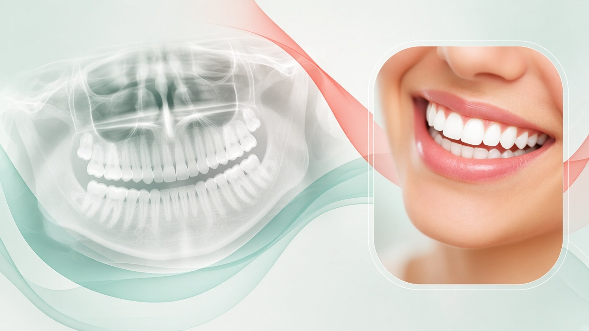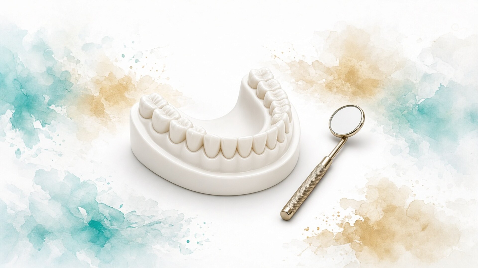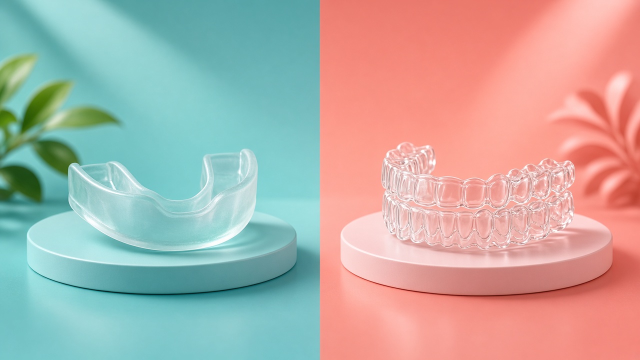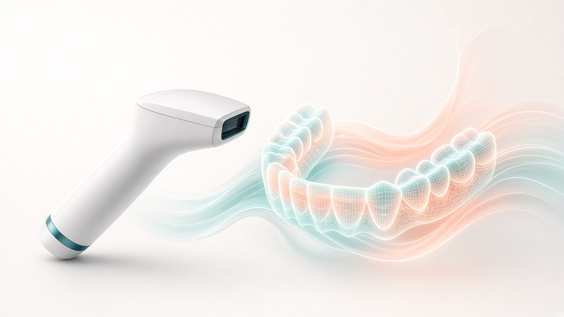
A digital dental scan has replaced almost every classic impression in my practice. I am Dr. Maksut Behruzoglu, a specialist orthodontist in Dubai, and I record your mouth with an intraoral scanner: a small camera on a wand captures thousands of images per second, and software stitches them into a 3D copy of your teeth, gums, and bite. Both arches plus a bite record take five to fifteen minutes in experienced hands. No tray of putty, no waiting for it to set, no gagging marathon. Studies from recent years agree that on short spans a scan matches silicone impression accuracy, while full-arch scans show slightly larger deviations that still sit well inside what orthodontics and clear aligners need. Scanning does not suit every case. With no remaining teeth, long bridges on several implants, heavy bleeding, or margins deep under the gum, I or my restorative colleagues still take a classic impression or combine methods. Below: how the scan works, what drives accuracy, where impressions still win, and what the model does for your treatment plan.
What happens during a scan appointment
Patients who remember childhood molds walk in tense: a tray of pink goop, material creeping toward the throat. A digital impression and 3D scan visit looks different. You sit in the chair while I hold a wand roughly the size of a thick toothbrush and glide it along your teeth. On the screen next to you, your teeth and gums appear in color, piece by piece. If the scanner misses a spot, the software shows a hole and I go back to it. You can stop at any second: raise a hand, I pull the wand out, you swallow and rest. Then we resume from the same point without losing the model. Talking during a scan is awkward, so we agree on the hand signal before we start. Below are the three stages of every scan.
Preparation: a dry field and good light
The scanner records surfaces optically. Saliva, blood, and glare bother it the way raindrops bother a photographer shooting through a window. Before we start, I ask you to rinse, my assistant places a saliva ejector, and sometimes cotton rolls or a cheek retractor. Plaque and food debris show up in the scan as part of the tooth, so brushing and skipping snacks for half an hour before the visit helps.
Metal fillings, crowns, and polished ceramics create glare. Current scanners handle this without powder. Older models needed a matting spray; today I use it rarely, on a single metallic surface here and there. Braces can be scanned too, though wires and ties cast shadows and the job takes longer.
The scan itself: one arch at a time
I start with the lower or upper arch depending on the task. The wand travels across the chewing surfaces, then the tongue side, then the cheek side. That sequence helps the software stitch frames cleanly. A full arch takes me one to three minutes; a newer operator needs more.
Your tongue and cheeks move, and that is fine. I may ask you to relax the tongue or shift it aside, or I hold a cheek back with a mirror. Breathe through your nose or mouth, whichever feels natural. The gag reflex fires less often than with a tray because the wand never covers the whole palate and never sits in one spot for minutes. If your reflex is strong, I scan the back teeth in short passes with breaks.
Bite registration and model check
After both arches I ask you to close in your usual bite. I scan from the left and right sides, and the software aligns the upper and lower models to that bite. If you close in more than one way, I ask you to repeat and watch that the position stays natural, without pushing the jaw forward.
Then I review the model on screen: gaps at tooth edges, the last molar, matching contacts on both sides. You see the model with me. I point out crowding, worn areas, spaces, and tipped teeth. For many people it is the first time they see their bite from the side and from behind. That conversation over the model often teaches more than ten minutes of hand-waving.
How accurate is a digital scan?
Accuracy is the first question engineers ask, and the right one. Every method carries some error. Researchers measure two things. Trueness tells you how closely the model matches the real teeth. Precision tells you how closely repeat scans of the same mouth match each other. Reviews published between 2020 and 2025 paint a consistent picture: for a single tooth, a quadrant, or a short bridge, intraoral scanners reach the accuracy of silicone impressions or better. Across a full arch, stitching error accumulates, and deviations of tens of microns appear more often. In orthodontics that error stays clinically invisible, because we move teeth fractions of a millimeter per stage and check aligner fit in the mouth. Crowns carry stricter demands, and there I defer to the restorative dentist. Below are the three factors that decide real-world accuracy.
Short span versus full arch
The scanner builds the model from many overlapping frames. Each join adds a tiny error. On three teeth there are few joins, and the errors have no room to add up. Across an arch from the last right molar to the last left molar there are hundreds, and a slight bend in the model becomes measurable.
For a single crown this barely matters. For a long framework that must seat on several implants at once, it matters a lot: a metal frame does not forgive distortion the way a plastic aligner does. That is why prosthodontists still debate methods for long-span work, while in orthodontics the debate has largely settled.
What undermines accuracy
The scanner’s main enemies are moisture, movement, and haste. Saliva on a tooth creates a false surface. Blood from inflamed gum hides the edge. If the operator moves the wand too fast or in jerks, the software loses reference points and stitches frames with errors. If you move your tongue while I capture the last molar, the end of the model drifts.
The second layer is the device and its software. Scanners from different generations differ in speed and capture quality, and software updates change results too. I keep my scanner on current software and check calibration on the manufacturer’s schedule.
The third layer is the clinician. The same scanner in two sets of hands produces two different models. Scan path, moisture control, and the habit of spotting a gap on screen come with hundreds of scans.
Accuracy for orthodontics and aligners
For an orthodontic plan I need crown shape and position, gum line, and bite. A scan delivers all of that with room to spare. Aligner manufacturers accept scans directly and build the virtual plan on them. In published comparisons, aligners made from scans fit at least as well as aligners made from silicone impressions.
Retainers come from scans as well. If you lose a retainer and your model sits in the archive, a lab can make a new one without another visit, as long as your teeth have not moved since. I check that separately, because an old file reflects an old tooth position.
The classic impression: how it works and where it shines
Impressions remain a working method, and I do not treat them as a relic. The principle is simple: the dentist fills a tray with soft material, seats it in your mouth, waits for it to set, and removes it. The result is a negative of your teeth, which a technician pours in stone to make a model.
There are two main material families. Alginate is a powder mixed with water. It is inexpensive, sets in a couple of minutes, and works for study models, simple appliances, and training. Its weak spot: alginate loses or absorbs water and changes shape if nobody pours the model soon. Drive an alginate impression across Dubai in August and accuracy suffers.
Silicone materials (addition silicone, often called PVS) are more stable. The dentist takes them in two layers or with two viscosities, and the lab can pour the model hours or even days later. Silicone captures fine detail: prepared tooth margins, gum contour around an implant. For crowns and bridges it served for decades as the benchmark against which scanners are measured.
Impressions outperform scanners in a few situations. They capture movable tissue in function: while you move your lips and cheeks, the material shapes the border of a future denture to your muscle activity. They do not depend on optics, so blood and saliva interfere with them in different ways. They need no electricity, license, or internet connection. Any lab in the world accepts a stone model.
Patients know the downsides firsthand. The material fills the mouth, and some people gag or panic. Errors appear only after removal: a bubble, a tear, a short pour near the last molar. Then the impression gets repeated. Stone models take up space, chip, and vanish during a move. Every new appliance needs a new impression or a carefully stored model.
Stone served my orthodontic work for years. On stone I learned to measure space deficiency, sum tooth widths, and plan extractions or expansion. A skilled technician makes a precise plate or retainer from a good stone model. When patients bring childhood models, I study them with real interest: they show the bite before the first treatment and help explain where a relapse came from.
My switch to scanning happened for practical reasons. Patients stopped dreading diagnostic visits. Aligners arrived sooner. The archive stopped filling a cupboard. I gained the ability to overlay models from different years and show change in numbers. Those reasons concern convenience and speed. A well-taken impression stays accurate, and I tell patients so plainly.
Repeats deserve a word. With an impression, a repeat means another load of material, five more minutes with a tray, and another round of fighting the reflex. With a scan, I rescan only the missed area: the gap on screen closes in twenty seconds while the rest of the model stays put. For anxious patients that difference outweighs any micron.
Scan and impression compared in one table
The table below is the working cheat sheet I keep in my head when choosing a method. Figures are averages from my practice and published work; timing and materials vary between clinics. I choose the method for your case after the exam.
| Factor | Alginate impression | Silicone (PVS) impression | Intraoral scan |
|---|---|---|---|
| Chair time, both arches plus bite | 10–15 minutes | 15–25 minutes | 5–15 minutes |
| Comfort and gag reflex | Fills the mouth, gagging common | Longer in the mouth, gagging common | Wand partly in the mouth, pause any time |
| Accuracy, short span | Adequate for study models | High, benchmark for crowns | High, comparable to silicone |
| Accuracy, full arch | Depends on how soon it is poured | High | Adequate for orthodontics, debated for long prosthetics |
| Stability before pouring or sending | Minutes, sensitive to moisture and heat | Hours to days | File does not change |
| Storage and copies | Stone, shelf space, chipping risk | Stone, shelf space, chipping risk | Archived file, copies in seconds |
| Sending to the lab | Courier | Courier | File upload the same day |
| When I choose it | Simple study models, backup option | No teeth, long implant frameworks, deep margins | Orthodontics, aligners, retainers, most crowns |
| Limitations | Distortion, bubbles, repeats | Time, material cost, discomfort | Moisture, blood, movable tissue, limited opening |
Both methods are alive, and each owns its territory. In orthodontics the scan covers nearly every task, so my patients rarely see a tray.
When I still take a classic impression
The honest answer I give patients is this: a scanner covers most of my work, yet in specific cases I or a colleague choose an impression or combine the two. The decision depends on what the technician needs to receive and on the conditions in your mouth that day. Orthodontics seldom runs into these limits because it works with tooth crowns and the bite. If a restorative colleague asks for a silicone impression for a long implant bridge, that colleague is making a professional call for a specific framework. I encourage you to ask why a method was chosen and to expect a concrete reason. A good clinician names it in a sentence or two and shows what they want the lab to receive. Below are four situations where the classic impression keeps its place.
No remaining teeth and full dentures
On a jaw with no teeth the scanner has little to grip: the gum surface is smooth and uniform, without clear landmarks, so stitching suffers. A full denture also rests on soft tissue that changes shape under pressure and as the lips and cheeks move. The prosthodontist shapes the denture border in function, asking you to smile, purse your lips, and swallow while the material sets. A scanner records tissue at rest and cannot take that functional imprint. Digital protocols for edentulous jaws keep improving, yet many prosthodontists combine them with an impression.
Long frameworks on several implants
When one framework must seat on four to six implants at once, full-arch stitching error can produce a misfit. Metal does not forgive misfit: the framework seats under strain and screws loosen. Here prosthodontists use splinted silicone impressions, dedicated scan bodies, or photogrammetry. As the orthodontist I consult on tooth position; the method choice stays with the prosthodontist.
Bleeding, deep margins, and limited opening
If the gum is inflamed and bleeding, optics cannot see a tooth edge under blood. If a crown margin sits deep below the gum, light does not reach it without good retraction. A silicone material, with proper preparation, displaces fluid and flows into the sulcus. In orthodontics I more often postpone the scan by a week after a professional cleaning than switch methods.
Limited mouth opening from a jaw joint problem or after surgery hampers both methods. The scanner wand usually passes more easily than a tray, but the last molars can stay out of reach. Then I scan in segments or take a partial impression.
Technical failure and compatibility
A scanner is a device. It can break, software can fail to update, and a cloud service can be down at the moment of upload. For an orthodontic record I simply reschedule the scan. For an urgent job, such as a broken retainer before a flight home, my assistant can take an alginate impression the same day.
The reverse happens less often: a lab abroad works only with stone or cannot read a file format. Files convert to a common format, and the lab can 3D-print a model. I confirm this before work begins.
What a scan adds to your orthodontic plan
For an orthodontist the dental model is the foundation of the plan. I used to work on stone: measuring tooth widths with calipers, sawing a duplicate model, resetting teeth in wax. Today software does that on a digital model, and the decisions stay with me. During aligner treatment planning I load the scan, stage the tooth movements, and see which moves are realistic, where extra space is needed, and where attachments or interproximal reduction come in. You see the simulation, and we discuss the goal on screen before payment and before the first aligner. The simulation shows the clinician’s intent. Bone, gum, and roots respond differently in each person, so I verify the result in the mouth at check-ups and adjust the plan along the way when needed. Below are the three practical benefits patients value most.
The model as a record of your starting point
A scan captures every tooth position on the day of the exam. A year into treatment I can overlay the new model on the old one and show, in millimeters, how far a tooth moved, how much the arch widened, how the bite changed. That used to require two stone models and a ruler.
For you it settles arguments with memory. People adapt to a new smile fast and forget where they started. The model and photos answer objectively. If treatment runs slower than planned, the overlay shows which tooth lags, and I change the plan based on facts.
Aligner plans and retainers
For aligners the scan has become standard. The manufacturer receives the file the day of the exam, I edit the virtual plan, you approve it, and the lab prints models and forms the trays. The chain runs shorter than with couriers and stone, with fewer chances for transport damage.
If you need additional aligner series mid-treatment, I rescan in the same visit. The new plan builds on where your teeth actually sit. Retainers at the end of treatment also come from a fresh scan taken on the finishing day, so they hold the achieved tooth position.
Comparing scans over time
After treatment, scans help track stability. At a six- or twelve-month check I take a new model and overlay it on the model from debond day or the last aligner. Small shifts show up before you notice them in the mirror.
Adults whose teeth started shifting after thirty often arrive asking how bad it is. If an old scan exists in an archive, the answer is precise. If not, we scan now, and today becomes the reference point for the future.
Comfort, children, hygiene, and safety
A scan uses no X-rays. The scanner works with visible light, like a camera with a built-in lamp. Pregnancy, children, and frequent repeats create no dose concerns. When radiographs are needed for planning, I order them separately and only when indicated.
Children tolerate scanning better than impressions. A child can watch the screen as their teeth appear and take breaks. An anxious child who once choked on alginate often settles within a minute. With young children I scan in short passes and do not demand perfect stillness; the software forgives small movements.
Adults with a strong gag reflex gain the most. I warn you where it may feel unpleasant, work from the front teeth backward, and pause often. Most people finish without an episode. If the reflex does fire, we stop, and the model does not need to start over.
People with claustrophobia or a hard dental history can hold the wand first, look at it, feel its weight. Then I make one short pass over the front teeth and show the result on screen. Once you see the process is controlled and stops on your signal, the rest usually goes calmly. That step costs two minutes and saves stress across the whole course, since a treatment usually involves several scans.
Wand hygiene follows the clinic’s infection-control standards: some tips go through the autoclave, some are single-use, and the body is disinfected between patients. If you want to know the protocol, ask the assistant; a good clinic answers without hesitation.
Temperature and taste matter to some people. Impression material is cold and has a smell and flavor. The scanner tip is heated to prevent fogging and leaves nothing in your mouth.
In Dubai orthodontic practices, the scan usually sits inside the diagnostic visit or the treatment package rather than appearing as a separate line. If cost affects your decision, ask the front desk before the visit and check the price-list date, because fees change.
Files, storage, and moving between countries
Dubai residents move often: a new job, a return home, treatment started in one country and finished in another. For them a digital model solves a practical problem. Stone gets lost in moving boxes; a file stays in the clinic archive and the manufacturer’s cloud.
Scans are saved in formats that labs and software read worldwide. If you relocate, I can send the model to your next orthodontist along with the plan, photos, and radiographs. That needs your request and consent to share data. The model is part of your medical record, and you have the right to receive it.
A file carries one limit worth remembering. It reflects your teeth on scan day. Six months without retention can shift teeth, and the old model will no longer fit a new aligner or retainer. I always check how fresh a model is before ordering an appliance from the archive.
Your data is protected the same way as the rest of your medical record. Scanner and aligner manufacturers store files on their servers under their own privacy policies. If the physical location of your data matters to you, ask before the scan.
Patients who switch to me from another orthodontist sometimes bring scans from abroad. I open them and check completeness and date. If the model reads well and the teeth have not moved since, I use it as a starting reference for comparison. For a new plan I still take a fresh scan, because the plan has to fit today’s mouth.
Cost and turnaround for appliances made from scans depend on the lab and manufacturer. On average, the path from scan to first aligners or a retainer runs shorter than from an impression, because courier trips and pouring drop out of the chain. I give an exact timeline after we choose the system and lab.
Frequently asked questions about digital dental scans
Does a digital dental scan hurt?
No, a scan causes no pain. The wand touches your teeth and cheeks, and I may move a cheek or your tongue aside, which can feel unfamiliar. There are no injections, no drilling, and no pressure on the teeth. If you feel uncomfortable, raise a hand and I stop.
How long does a scan take?
In experienced hands, both arches plus the bite take five to fifteen minutes. Timing depends on the number of teeth, braces, saliva flow, and your gag reflex. A first scan at a new clinic runs longer because I explain the model as I go. Repeat scans at check-ups move faster.
Is a scan more accurate than an impression?
On short spans, scan accuracy matches silicone impressions; across a full arch the error runs slightly higher. For orthodontics, aligners, and retainers, a scan is accurate with room to spare. For long frameworks on several implants and full dentures, clinicians often choose silicone or combine methods. The right method depends on the job.
Can you scan my teeth with braces on?
Yes. Brackets and wires cast shadows, so the scan takes longer and I need more passes from different angles. A model with braces works for tracking tooth movement and planning the next stage. For retainers after braces come off, I take a new scan without them.
Is scanning safe during pregnancy or for children?
A scan uses visible light and involves no X-rays. There is no dose limit, so it can be repeated as often as treatment requires. For pregnant patients I adjust the chair position and add breaks if nausea is an issue. Children usually find a scan easier than an impression.
Can I get a copy of my scan file?
Yes, the model is part of your medical record. On request I send the file to you or to your next clinician in a format labs can read. Keep in mind that the file reflects your teeth on scan day. If a lot of time has passed, a new clinician will need a fresh model.
Do I need a scan if I already have stone models?
Stone models help as a starting reference and for comparison. For a new plan, especially with aligners, I need a fresh digital model because your teeth may have moved since the stone was poured. A lab can also scan old stone models to keep them in a digital archive. I decide which old records are useful after the exam.
Why did another clinic take an impression instead of a scan?
There are several reasons, and most say nothing bad about the clinic. The dentist may have chosen silicone for a job where it performs better, such as a long implant denture. The scanner may have been booked or under repair. Ask about the reason; a good clinician explains it clearly.