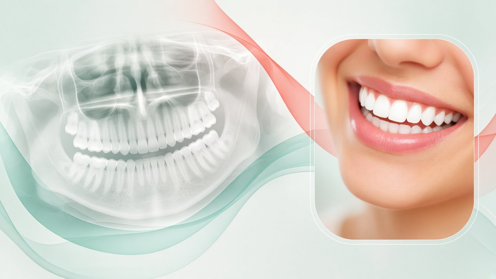
A dental CT scan, which in a dental setting almost always means CBCT (cone-beam computed tomography), delivers a dose you can measure and compare with everyday exposures. I am Dr. Maksut Behruzoglu, a specialist orthodontist in Dubai, and in X-ray and CT imaging visits I give patients the numbers. A periapical X-ray is usually 1–5 microsieverts, a panoramic (OPG) about 5–25, a small-field CBCT roughly 10–100, and a full-face CBCT from 50 to several hundred, higher on some units. For context, average natural background radiation worldwide is about 2.4 millisieverts a year, roughly 6–7 microsieverts a day. A hospital CT of the jaws typically delivers several times more than dental CBCT. The spread depends on the machine, field of view, settings and age. Low doses do not mean zero risk, so I follow the ALARA principle: a scan is ordered when the answer will change treatment, and taken with the smallest field and dose that still answers the question. In pregnancy I postpone routine imaging; urgent scans go ahead when needed. Below: the numbers, what drives them, and how to decide calmly.
Why dental CT scans worry people
The word "tomography" sounds serious. Many people link it with hospitals, a worrying diagnosis, a tunnel-shaped machine and a doctor saying "we need a closer look." When a dentist or orthodontist suggests a CT scan, a patient hears "radiation" and recalls everything they have ever read about it.
The second source of worry is terminology. "Dental CT" can refer to two different technologies. Multi-slice CT (MSCT, sometimes called medical CT) is the hospital scanner you lie down in, with a rotating ring. Cone-beam CT is the unit found in dental clinics and imaging centres; you stand or sit, and the machine makes a single turn around your head. For teeth and jaws, dentistry uses CBCT almost exclusively. Its dose is usually lower than MSCT of the same area, sometimes by several times.
The third source is numbers without context. A patient reads that CBCT gives "dozens of times more radiation than a panoramic," and for some settings that is true. Dozens of times a very small number is still a small number. Without comparisons to natural background, a flight, or a chest X-ray, lines like that only frighten.
I do not brush this worry aside. Radiation without benefit is pointless, and a good clinician should explain why a scan is being ordered. Waving it away with "it's nothing, everyone has one" is wrong too: low doses carry a small but non-zero risk, particularly in children. My job is to give you the numbers, the context and the reasoning. We make the decision about the scan together.
How radiation dose is measured and compared
To compare different exams, radiologists use effective dose, measured in sieverts. It accounts for the amount of radiation and for which organs were in the beam and how sensitive they are. The thyroid, salivary glands, jaw bone marrow and eye lens each carry their own weighting, and effective dose combines them into one figure. In dentistry the handy unit is the microsievert, one millionth of a sievert. The figures you see in articles are nearly always estimates measured on phantoms, mannequins fitted with sensors, rather than on a specific person. Two identical units with different settings can differ several-fold. So I give patients a range and explain what moves the number up or down. Below: what a microsievert means, how it compares with daily life, and why sources disagree.
Microsieverts in plain terms
One sievert is a dose at which the risk of long-term effects, mainly cancer, becomes noticeable across a population. Dentistry never comes near that. A millisievert is a thousand times smaller, a microsievert a million times smaller.
Dental X-rays are measured in microsieverts: single digits, tens, occasionally hundreds. A medical CT of the head or chest is measured in millisieverts. The gap is tenfold or more.
To estimate low-dose risk, radiation protection uses a cautious linear model: every dose is assumed to add a proportionally small risk. The International Commission on Radiological Protection (ICRP) uses a population coefficient of about 5% per sievert. Plug in 100 microsieverts and you get an added lifetime risk on the order of one case per 200,000 people. Many experts think the model overstates risk at very low doses, but it is used as a conservative safeguard. For children the coefficient is two to three times higher.
Background radiation and flights
Everyone is exposed all the time: cosmic rays, radon in indoor air, rocks and soil, even the potassium in your own body. The global average is about 2.4 millisieverts a year, roughly 6–7 microsieverts a day. Some regions of the world have background levels several times higher, and people have lived there for generations.
Flights add cosmic radiation at altitude. At cruising height a passenger receives roughly 3–5 microsieverts an hour, depending on route latitude and solar activity. A seven-hour Dubai–London flight gives about 20–40 microsieverts. A flight to Istanbul or Moscow is less, around 10–25.
These comparisons make the scale tangible. A panoramic X-ray equals a few days of ordinary life on Earth or a few hours in the air. A small-field CBCT equals one to two weeks of background or one long-haul flight. A large-field CBCT on some machines can reach a month or two of background.
Why the same exam has different numbers
Patients find CBCT figures online ranging from 10 to 1,000 microsieverts and cannot tell which to trust. Both ends of the range are correct; they describe different situations.
The European SEDENTEXCT project, which compiled data on dental CBCT, found dose differences of more than tenfold between units and settings. A small field over two teeth in low-dose mode and a full-face field at high resolution on an older unit differ like a bicycle and a lorry.
Patient age also shifts effective dose: in a child the organs sit closer together and the thyroid is more likely to fall inside the beam. Finally, calculation methods have changed. In 2007 the ICRP revised its tissue weightings, including salivary glands, and figures calculated before that revision run systematically lower. Only numbers calculated by the same method can be compared fairly.
Dental imaging doses in one table
The figures below are approximate, drawn from reviews and European guidance of recent years. They describe typical ranges for modern digital units in adults. Your actual dose depends on the unit, settings, field of view and age, and the imaging centre can tell you the figure for its machine. Background equivalents use a global average of about 6.5 microsieverts per day.
| Exam | Typical effective dose, µSv | Natural background equivalent | Comparable flight time | Biggest dose driver | Typical orthodontic use |
|---|---|---|---|---|---|
| Periapical X-ray | 1–5 | a few hours to 1 day | about 1 hour | Detector, collimation | Root, root tip, doubtful tooth |
| OPG (panoramic) | 5–25 | 1–4 days | 1–6 hours | Unit, exposure program | Start of treatment, teeth and bone overview |
| Lateral ceph | 2–10 | up to 1.5 days | 1–3 hours | Collimation, thyroid-area filter | Skeletal class, growth, profile |
| Chest X-ray (for comparison) | about 20 | about 3 days | about 5 hours | Unit, projection | Not orthodontic |
| Small-field CBCT (1–3 teeth, one joint) | 10–100 | 1.5 days to 2 weeks | 3 hours to one or two long-haul flights | Field size, voxel, mode | Impacted canine, neighbouring roots |
| Medium-field CBCT (one or both jaws) | 30–250 | 5 days to 5 weeks | a short flight to several long-haul flights | Field, mode, unit | Tricky mini-screw sites, adult expansion |
| Large-field CBCT (full face) | 50–600, higher on some units | 1 week to 3 months | several long-haul flights | High-resolution mode, unit age | Jaw surgery, marked asymmetry |
| Hospital MSCT of jaws (for comparison) | 400–2000 | 2 to 10 months | dozens of flight hours | Protocol, slice count | Rarely used in orthodontics |
The table shows the main point: dental imaging sits in the low-dose zone, and the differences between methods are real. A large-field CBCT can deliver dozens of times more than a panoramic. That is why I do not swap flat images for volume without a reason, and I do not avoid volume when the case calls for it.
What drives CBCT dose
Two patients who both "had a dental CT" may have received doses ten or more times apart. That is neither a mistake nor carelessness at one centre, as long as each setting was chosen for its task. CBCT dose depends on several parameters the clinician and radiographer set before the scan. The biggest one is the field of view, meaning how much anatomy the beam covers. The second is the exposure mode: tube current and voltage, exposure time, the number of projections per rotation and the voxel size. The third is the unit itself: detector generation, beam filtration, and whether it offers low-dose and paediatric protocols. Age and head position matter too. When I write a referral, I state the question and the area so the centre can choose the lowest settings for the task. Below, each factor in turn.
Field of view
Field of view has the largest effect. A small field, around 4–5 cm, covers a few teeth or one joint. The thyroid, eye lenses and most of the salivary glands stay outside the beam. A large field, 15–20 cm or more, covers the entire face, and far more sensitive organs fall inside.
For an impacted canine I need the canine and the neighbouring incisors, not the whole face. For planning jaw surgery I need the full face, and a small field will not help.
Sometimes one medium field beats two small ones. If both upper canines are impacted, a single field over the front of the upper jaw usually gives a lower total dose than two separate scans.
Resolution and exposure mode
Voxel size sets the level of detail. A voxel of about 0.1 mm shows fine structures: early root resorption, a crack, a thin cortical plate. A 0.3–0.4 mm voxel gives a coarser image that is often enough to locate a tooth or assess bone volume.
A smaller voxel usually needs more radiation to keep image noise down. So I request high-resolution mode only when the question involves fine structures. Locating an impacted tooth often works in standard mode.
Many modern units offer low-dose modes with fewer projections or lower tube current. Image quality drops, but for some questions it is sufficient. A check on the position of a canine after it has been brought into the arch, for example, can often be done that way.
Unit, age and repeat scans
Newer units with sensitive detectors and good filtration deliver less dose at similar image quality. An imaging centre can tell you the dose for a given mode on its machine, and asking is perfectly reasonable.
Age affects risk more than the dose number itself. A child's tissues are more sensitive and there are more years ahead for a theoretical long-term effect to appear. So I order CBCT for children less often and request a paediatric protocol.
Repeat scans add up. If within a year a patient has a CBCT with an implant dentist, a periodontist and an orthodontist, the total rises even though each scan was justified on its own. I ask patients to bring every recent scan, and often I can work from an existing volume.
The ALARA principle: how I decide whether a scan is needed
ALARA stands for "as low as reasonably achievable." Dentistry often adds ALADA, "as low as diagnostically acceptable": the dose should be the minimum at which the image still answers the clinical question. In practice the principle works in three steps. First, justification: will the scan help a decision? Second, optimisation: how to get the answer with the least dose. Third, an honest look at the other side, because skipping a needed scan carries its own risk. The American Association of Orthodontists (AAO), the American Dental Association (ADA) and European dental radiology guidelines describe these steps in similar terms. I run through them at every orthodontist consultation before I write any imaging referral. Below, each step with examples from my practice.
Justification
Before ordering a scan I ask myself one question: will the answer change the treatment plan? If a panoramic has already shown everything I need, a "just in case" CBCT adds nothing. If a ceph will help me choose between a functional appliance and braces for a teenager, it is justified.
Routine CBCT for every orthodontic patient is not part of international guidance. Volume imaging belongs to specific questions: an impacted tooth that flat images cannot locate, suspected root resorption, marked asymmetry, mini-screws in a tricky site, surgical planning.
I write the question the scan must answer on the referral. That keeps both me and the imaging centre disciplined: a scan without a question is hard to read, and a scan with one is read quickly and precisely.
Optimisation
Once a scan is justified, the next task is getting the answer at the lowest dose. I choose the smallest field, a standard voxel when fine detail is not needed, and low-dose mode where it fits.
Part of optimisation is reusing what already exists. If a patient had a CBCT six months ago for an implant dentist and it covers the area I need, I work from it. A volume can produce a panoramic reconstruction and a synthetic ceph, so separate flat images become unnecessary.
Another part is positioning. If a patient moves during the scan or stands incorrectly, the image blurs and has to be repeated, which doubles the dose. So I explain in advance: stay still for about 10–20 seconds, do not swallow, breathe calmly.
When skipping a scan is also a risk
Some patients decline a justified scan because they fear radiation. I respect that decision, and I show what sits on the other side of the scale.
An impacted canine without CBCT means the surgeon exposes the tooth without knowing exactly which side it lies on, and I pull it without seeing how it relates to the lateral incisor root. Resorption of the neighbouring root may then be found late, once the root has already shortened noticeably. Losing an incisor at twenty means an implant and decades of follow-up care.
A mini-screw placed blind into a tight space between roots can hit a root. Expanding an adult's upper jaw without checking the cheek-side bone can end in gum recession. Those risks are real, and next to them a small-field CBCT dose comparable to a few days of background looks different. The decision stays with the patient; my job is to make sure both sides are visible.
Pregnancy and dental X-rays
Pregnancy is its own topic, and I ask patients to tell me about a pregnancy, or a possible one, before any scan, even if it is very early and not yet confirmed. That information decides which images we postpone. The reassuring part is that dental imaging targets the head, and the baby is far from the beam. Scattered radiation reaching the abdomen during a jaw scan is estimated to be orders of magnitude below levels at which any effect on the fetus is discussed. The American College of Obstetricians and Gynecologists (ACOG) states that doses below 50 milligray have not been linked with developmental anomalies, and dental imaging gives the fetus a tiny fraction of that. Pregnancy still reshuffles priorities, and orthodontic treatment is usually not started during this period. Below: routine scans, urgent situations and shielding.
Routine scans can wait
Orthodontic treatment is rarely urgent. If you are pregnant and want to start straightening your teeth, I usually suggest postponing the start, and the scans that go with it, until after the birth. There are several reasons: hormones change gum health, visits and new appliances add strain, and there is no rush.
If you become pregnant during treatment, we carry on, but new routine scans wait. A check panoramic before braces come off can be done after delivery unless there is a specific warning sign.
A consultation without imaging can happen at any stage: exam, photos, scan, plan discussion. It involves no radiation and helps you prepare.
Urgent situations
Acute tooth pain, swelling, trauma or suspected infection cannot wait for the end of pregnancy. Infection and pain carry more risk for mother and baby than a periapical or panoramic X-ray. In those cases the dentist or surgeon takes the image needed for a decision, which is consistent with ADA and obstetric guidance.
CBCT during pregnancy is rare and reserved for urgent questions that flat images cannot answer, with the smallest field and mode. The clinician managing the emergency makes that call, together with your obstetrician when needed.
If you find out you were pregnant after a scan taken before you knew, there is no reason to panic. Dental imaging doses to the fetus do not come close to levels at which intervention is discussed. Let your obstetrician know so it goes in your record.
Lead aprons, thyroid collars and breastfeeding
Many people expect a lead apron for every X-ray. In 2024 the ADA updated its recommendations and no longer advises routine lead aprons or thyroid collars for dental imaging. The reasoning: modern units collimate the beam tightly, the apron's protective contribution is minimal, and a collar can intrude into the image and force a repeat.
Rules vary between countries and clinics. Many centres in Dubai still offer an apron, especially to pregnant patients. If an apron makes you feel better, ask for one; it is a reasonable request and in most cases does not interfere with the image.
There is no need to pause breastfeeding after a dental X-ray or CBCT. X-rays do not stay in the body and do not reach breast milk. Dental CBCT uses no contrast agent, so feeding carries no restriction.
Children and teenagers: why extra caution applies
Most of my patients who need orthodontic imaging are children and teenagers, and for them the dose question matters more. Growing tissues are more sensitive, a child's thyroid sits closer to the beam, and there are more years ahead for a theoretical long-term risk to show. By standard estimates, the same scan carries two to three times the calculated risk in a child compared with an adult.
So I follow a few rules for children. First, flat images when they answer the question. Panoramic and ceph cover most tasks in children and teens: developing teeth, the change from baby to adult teeth, jaw relationships, growth. Second, CBCT only for a specific indication, most often an impacted canine, extra teeth between the incisors, or trauma. Third, the smallest field and a paediatric protocol if the unit supports one. The international Image Gently campaign for paediatric imaging promotes the same approach.
Parents sometimes ask whether it would be better to do a CBCT straight away "so we don't have to repeat it." The logic is understandable, but children change fast. A CBCT at eight will not show where the canine will be at twelve. A scan taken too early often has to be repeated anyway. I order volume imaging at the point where a decision depends on it.
The opposite also happens. Parents turn down a panoramic for an eight- or nine-year-old because "it's too early." At that age the film shows whether all adult tooth buds are present and whether there are extra teeth. A missed case of absent premolars or an extra tooth between the incisors costs far more than a panoramic dose equal to a couple of days of background.
I keep a log of each child's imaging. If a general dentist took a panoramic within the past year, I ask for it rather than ordering a new one without cause. I advise parents to keep files of every scan, especially if the family moves often. In Dubai that story comes up regularly: the images stayed at a clinic in another country, and without them we have to repeat.
How to prepare for a CBCT and what to ask
Preparation is simple, and it determines whether the scan has to be repeated. Before the scan, remove earrings, facial and tongue piercings, glasses, removable dentures and retainers, and hair clips. Metal in the field creates artefacts, bright streaks that hide nearby structures. Fixed braces stay on; the radiologist accounts for their artefacts, and for most orthodontic questions the image remains readable.
During the scan the unit rotates around your head for 5 to 40 seconds, depending on the mode. Your job is to stand or sit still, not swallow and not talk. The radiographer positions your head with a chin rest and supports. If you are prone to claustrophobia, CBCT is usually easy to tolerate: the unit is open, with no tunnel.
Asking a few questions before the scan is completely normal. Which clinical question should this scan answer? What field of view was chosen and why? Can my recent scan be used instead of a new one? What dose does this mode give on your unit? Will I receive the files in DICOM format? A good clinician and a good centre answer these calmly.
After the scan, ask for the complete file set, not just the report or screenshots. DICOM files let another doctor review the volume without a repeat scan if you change clinic or country. That is a direct dose saving later on.
If you are unsure whether you need a CBCT, raise it at an orthodontist consultation. I will show you the question at stake, what the flat images already reveal, and what a volume scan could change. Sometimes after that talk we decide to go without CBCT; sometimes the patient agrees once they see what is at stake.
CBCT cost in Dubai depends on the centre, the field of view and your policy. If price comes up, I give an AED range with the price-list date and remind you to confirm coverage with your insurer, because orthodontic imaging is not covered the same way across plans.
Frequently asked questions about dental CT radiation
Is a dental CT scan harmful if I have one every year?
A single small- or medium-field CBCT dose is comparable to weeks of natural background, and its calculated risk is very small. I judge each scan on its own justification. If every scan answers a question that shapes treatment, a yearly CBCT can be reasonable. If scans are taken "to keep an eye on things" without a question, I suggest skipping them.
Which is safer, CBCT or a hospital CT?
For teeth and jaws, dental CBCT usually delivers a lower dose than multi-slice CT of the same area, sometimes by several times. CBCT also gives fine bone and tooth detail. Hospital CT shows soft tissue better and serves other purposes. For orthodontic questions CBCT is almost always enough.
Can I have a dental CT scan while pregnant?
I postpone routine orthodontic imaging until after pregnancy. In an urgent situation such as pain, swelling or trauma, the needed image goes ahead, because infection and pain carry greater risk. The fetal dose from dental imaging is orders of magnitude below levels at which any effect is discussed. Always mention a pregnancy before a scan.
Do I need to "flush out" radiation after a CT scan?
No. X-rays do not remain in the body after the exposure and do not build up in tissue like a substance. Special foods, pills or treatments to "remove radiation" after a CBCT are unnecessary and do nothing. Breastfeeding can continue without a break.
Why does one centre quote 30 microsieverts for CBCT and another 300?
The difference comes from field of view, exposure mode, voxel size and unit generation. A small field in low-dose mode and a large field at high resolution differ several-fold. Both figures can be correct for their task. Ask the centre which field and mode they chose and why.
Do I need a lead apron for a dental CT?
Current ADA recommendations no longer call for routine aprons or thyroid collars: the beam is tightly aimed and a collar can spoil the image. Clinic rules vary by country, and many Dubai centres offer an apron on request. If it makes you more comfortable, ask for one. Many centres use an apron by default for pregnant patients.
How many X-rays are safe for a child during orthodontic treatment?
There is no fixed limit; each scan is weighed on its justification. A typical course includes a panoramic, one or two cephs and, less often, a small-field CBCT for a specific reason. The combined dose of that set is comparable to a few weeks of natural background. I keep a record and do not repeat images without cause.
Can I skip CBCT and be treated from a panoramic?
In many cases yes, if the panoramic and ceph answer the planning questions. With impacted teeth, suspected root resorption or surgical planning, skipping CBCT raises clinical risk. I will explain what we cannot see without volume imaging and what that could lead to. The decision is yours once you have seen both sides.