Before braces or aligners I collect diagnostic records: 8 to 10 photos of the face and teeth, an intraoral scan, a panoramic X-ray (OPG), a lateral cephalogram and, for specific questions, a 3D scan (CBCT). Patients ask why an orthodontist needs X-rays when crooked teeth are plain to see. The eye sees crowns. It cannot see roots, bone, developing teeth, or the jaw relationship in degrees and millimetres, and those decide how far a tooth can move without harm. Modern 2D dental X-rays deliver a dose of a few to a few dozen microsieverts, about the same as several days of natural background radiation. CBCT delivers more, so I order it only to answer a defined question. Photos and scans involve no radiation at all. If you bring recent, good-quality films from another clinic, I use them instead of repeating the exposure. Below: what each record shows, which decisions depend on it, how the dose compares with everyday life, and when repeat X-rays during treatment make sense.
Why a visual exam is not enough for a plan
At the exam I see about a third of what I need for a plan. Crowns, gums, the bite from the front and sides, jaw movement. That is enough to say "treatment is likely indicated" or "we can watch this." It is not enough to calculate where each tooth should go.
A tooth sits in bone by its root, and the root is one and a half to two times longer than the visible crown. A crown can look straight while its root presses against a thin wall of bone. If I moved that tooth blind, I would risk gum recession or an exposed root. A panoramic X-ray, and a CBCT where needed, shows where the root sits and how much bone surrounds it.
The second layer is the jaws. Two patients with equally prominent upper teeth can have different causes. In one, the upper jaw sits forward. In another, the lower jaw sits back. In a third, the jaws are fine and the teeth are tipped. Treatment differs for each: a growing child in the first group may need a growth appliance, an adult in the second may need to discuss surgery, and braces alone may solve the third. Nobody can tell these apart by eye with confidence. A lateral cephalogram and a set of angle measurements can.
The third layer is what stays invisible. An impacted canine in the bone, an extra tooth, a missing permanent tooth bud, a cyst, short roots after a childhood injury. These findings turn up in a noticeable share of patients, and I do not want to discover them six months into treatment when a tooth refuses to move.
The fourth layer is a reference point. Photos and a scan taken before treatment record where you started. A year later we compare before and after images side by side. That matters for judging progress, for deciding on plan changes, and for protecting both sides if questions arise.
The fifth layer is teamwork. Adult orthodontics often runs alongside a general dentist, a periodontist, a surgeon or a restorative dentist. An implant surgeon wants to know where neighbouring roots will sit after alignment. A periodontist compares bone levels before and after. A surgeon plans an operation from the same images I use. One shared set of records lets everyone work from the same data, and spares you a separate round of X-rays at each specialist.
I explain all this at the first consultation, because distrust of X-rays is common. Some people have heard that clinics order extra imaging to pad the bill. Others fear radiation. Both concerns make sense, and I answer both with specifics: I order only what changes the plan, and I give you the dose in numbers.
The photo series: which shots and what they show
A standard orthodontic photo series is the first and simplest part of the records. It involves no radiation, takes five to ten minutes, and gives me material for analysing the face and smile that no X-ray can provide. Orthodontic associations describe a standard set: several face shots and five intraoral shots. I take the same set for every patient so that images before, during and after treatment share the same light and angle. I use a DSLR with a ring flash; a phone distorts too much at close range. Intraoral shots need plastic lip retractors and mirrors. They feel strange but do not hurt. The photos stay in your medical file and are never published without separate written consent. I repeat the series midway and at the end. Each group of shots answers a different question.
Face from the front and in profile
A face photo at rest shows symmetry, the balance of the upper, middle and lower thirds, and lip position. I check whether the lips close without straining the chin muscle. If you have to strain to close your lips, that points to protruding teeth or a vertical imbalance.
The profile shows facial convexity, chin position, and the angle between the nose and upper lip. That angle feeds into the extraction decision: with an acute angle and a full profile, extractions can improve the face; with an obtuse angle and a flat profile, they can make it worse. I never decide on one photo, but I never decide without it either.
For children I repeat profile photos every six to twelve months. Jaw growth shows up better across a series of images than at any single exam.
Smile and gum display
A smile photo shows how much tooth and gum you display, whether the dental midline matches the facial midline, and whether the line of the front teeth runs parallel to the eyes. These points shape how other people read your smile.
Patients with a "gummy smile" often want it corrected. The cause varies: excess vertical growth of the upper jaw, a short or very mobile upper lip, or teeth that have not fully erupted. Photos at rest and smiling help me tell one cause from another and pick the method: orthodontics, mini-screws, a periodontist's help, sometimes surgery.
I take a three-quarter smile to judge smile width and the dark triangles at the corners of the mouth. Those triangles sometimes shrink when the arch is widened.
Intraoral shots with mirrors
Inside the mouth I take five shots: teeth together from the front, right and left, plus the biting surfaces of the upper and lower arches through a mirror. The side shots show how the canines and molars meet, which determines the bite classification. The occlusal shots show arch shape, rotations and crowding.
These photos capture details a scan renders differently: enamel colour, white spots, fillings, gum condition. If white spots existed before braces, I record them. Two years later nobody has to argue about whether the braces caused them.
Sometimes I add a side view of the front tooth overlap or an open-mouth shot, depending on the case.
2D X-rays: OPG, cephalogram and periapicals
Two-dimensional X-rays remain the backbone of orthodontic diagnosis. They are fast, widely available and low-dose. The exposure itself lasts under a minute, and preparation means removing earrings, glasses and any removable dentures. Most dental clinics and all imaging centres in Dubai offer them. I order them after the exam, once I know which question needs answering. Most adults need a panoramic X-ray and a lateral cephalogram before braces. Children need a panoramic to assess developing teeth; I add a cephalogram if I suspect a jaw growth problem. Periapicals come in when a single tooth raises doubts. If you already have recent, good-quality films, I work from them. I write each referral for X-ray and CBCT imaging with a specific question so the radiographer knows what to look for. One 2D image cannot stand in for another.
Panoramic X-ray (OPG)
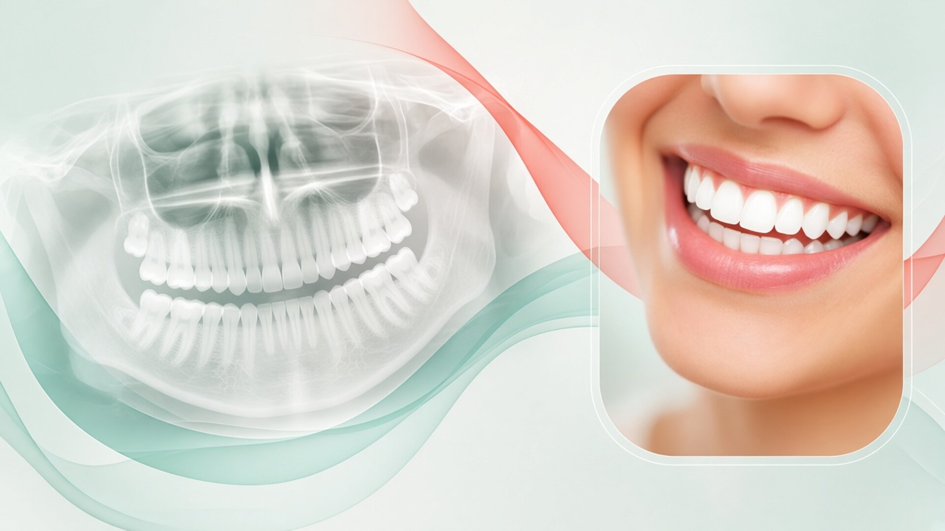
A panoramic X-ray shows every upper and lower tooth in one image, along with roots, developing teeth, the jawbones, the maxillary sinuses and the jaw joint heads. For an orthodontist it is the overview map.
On an OPG I check the number of teeth and tooth buds, extra or missing teeth, where the wisdom teeth are and how they are growing, root length, infection around root tips, and bone levels in adults. If a tooth has a short root after an old injury, I plan gentler forces for it. If a wisdom tooth grows at an angle into its neighbour, we discuss it before treatment starts.
A panoramic image distorts size and blurs the front teeth through overlap. It cannot give precise angles or the thickness of bone in front of and behind a root. Other methods answer those questions.
Lateral cephalogram
A lateral cephalogram shows the skull from the side: the cranial base, upper and lower jaws, teeth and the soft tissue profile. I mark reference points on the image and measure angles and distances. This process is called cephalometric analysis.
The analysis answers questions that shape the whole plan. How the jaws sit relative to the cranial base and to each other. How far the front teeth are tipped. Which facial growth pattern you have: horizontal, vertical or average. The answers decide whether tipping teeth will solve the problem, whether extractions are needed, whether a growth appliance makes sense for a teenager, and whether surgery belongs in the conversation for an adult.
Orthodontists also use the cephalogram to judge growth stage from the neck vertebrae. In a teenager that helps time a functional appliance. A repeat cephalogram at the end of treatment shows what we achieved in measurable terms.
Periapical X-rays of single teeth
A periapical X-ray shows one to three teeth in fine detail. I order one when something on the panoramic looks doubtful: a shadow at a root tip, suspected root shortening, decay under a filling, a crack.
Periapicals are useful during treatment too. If a tooth moves slower than expected or becomes mobile, a single-tooth image answers the question with a minimal dose. A full panoramic for one tooth is unnecessary.
For decay checks, general dentists use bitewing X-rays. I ask your dentist for them if I have doubts about your teeth before braces go on.
CBCT: when a 3D scan is worth it
Cone beam computed tomography shows teeth and bone in three dimensions. I can rotate the image, slice through any root and measure bone thickness to a tenth of a millimetre. The trade-off is a higher dose than 2D X-rays, depending on field of view and settings. The American Academy of Oral and Maxillofacial Radiology (AAOMR) and the European Orthodontic Society share a position: CBCT is justified when 2D images cannot answer the clinical question and the answer will change the plan. "CBCT for everyone before braces" does not fit that position. I order a 3D scan in roughly a third of cases, more often for adults and complex problems. I choose the smallest field of view that covers the question: one canine does not need a full-skull scan. The main indications and limits follow.
Impacted and displaced teeth
The most common reason for CBCT in orthodontics is an unerupted canine. A panoramic shows the canine in the bone at an angle. It does not show which side it lies on, palate or lip, or whether it has damaged the roots of the neighbouring incisors. CBCT answers both.
The answer drives the approach. A surgeon exposes a palatal canine differently from a labial one. If the canine has already resorbed an incisor root, the plan changes: sometimes we keep the incisor, sometimes the canine. A decision without 3D imaging is close to operating by guesswork.
The same applies to extra teeth, fused roots, and wisdom teeth near the nerve. The surgeon who will remove or expose the tooth often requests a CBCT anyway.
Adult bone and mini-screws
Adults have thinner bone around the roots than teenagers, especially at the front of the lower jaw. If the plan involves tipping or advancing the incisors, I want to know how much bone is there. A root pushed outside the bone risks gum recession, which is harder to fix afterwards.
Mini-screws, small temporary anchors for moving teeth, go between roots. If the space between roots is tight or a sinus sits close, a 3D scan helps pick a safe spot. In simple cases a panoramic and a clinical assessment are enough.
Before expanding the upper jaw in an adult, CBCT shows the state of the midline suture and bone thickness. That guides the choice of appliance: a conventional expander, a mini-screw-supported expander, or surgically assisted expansion.
What CBCT cannot show
CBCT shows bone and teeth well. It shows soft tissue, the jaw joint disc, muscles and nerves poorly. The joint disc needs an MRI.
Patients sometimes expect a 3D scan to answer questions about breathing. The airway is visible on CBCT, but the scan captures one posture while you are awake, and it cannot diagnose sleep apnea. That takes a sleep study with a sleep physician.
Metal crowns and old fillings create artefacts on CBCT. If you have a lot of metal in your mouth, part of the image will be distorted, and I account for that when reading it.
Radiation dose: the numbers, without fear
Fear of radiation is the main reason patients refuse X-rays or ask for "only the essentials." I take that fear seriously and answer with numbers. Everyone receives natural radiation from soil, air, space and food. The world average is about 2.4 to 3 millisieverts a year, or 7 to 8 microsieverts a day. Dental X-rays sit in the same range. The "as low as reasonably achievable" principle still applies: an X-ray without a reason is not needed, however small the dose. The figures below are approximate. Actual dose depends on the machine, settings and field of view, and your imaging centre can give exact values. I weigh the benefit to your plan against the dose before ordering anything. If an image will not change the plan, we skip it. Patients ask about three topics most often.
Compared with background radiation and flights
A periapical of one tooth delivers roughly 1 to 5 microsieverts, less than a day of background radiation. A panoramic on a modern digital unit delivers about 5 to 25 microsieverts, one to three days of background. A lateral cephalogram delivers about 2 to 6 microsieverts.
A small-field CBCT delivers around 20 to 100 microsieverts; a large-field scan ranges from 50 to several hundred, depending on the machine. That equals several days to several weeks of background radiation.
For comparison, a flight from Dubai to London takes about seven hours, and at cruising altitude a passenger receives a few dozen microsieverts. Frequent flyers in this city receive more from their travel than from a full course of orthodontic X-rays.
Children and teenagers
Children are more sensitive to radiation than adults: their tissues are growing and they have more years ahead. So I am stricter with imaging for children. I order a panoramic when I need to assess developing teeth and the change from baby to adult teeth, usually not before age 7 or 8. A cephalogram comes in if I suspect a skeletal problem and plan a growth appliance.
For a child, CBCT is reserved for a specific question, most often an impacted canine or an extra tooth, and uses the smallest field of view. Many children's consultations need no X-rays at all: an exam, photos and observation give enough information.
Parents ask how many X-rays a course involves. Typically a set at the start, sometimes one check midway, and one at the end. The exact number depends on the goal, and I list it in the plan.
Pregnancy and lead aprons
Orthodontics is elective care, so during pregnancy I postpone X-rays and the start of treatment unless there is an urgent reason. If you are planning a pregnancy, mention it at the consultation and we will choose the best order for X-rays and appliance fitting.
Many patients assume lead aprons and thyroid collars are mandatory. In 2023 the American Dental Association (ADA) revised its guidance: with modern equipment and proper beam collimation, aprons and thyroid collars are no longer recommended as routine, because they barely reduce dose and can block part of the image. Rules differ between countries and clinics. If you feel more comfortable with an apron, tell the radiographer.
If you are unsure whether you are pregnant, postpone the X-ray by a few days. It is a simple rule, and I ask every patient to follow it.
All diagnostic records in one table
To keep everything in one place, I have collected the records in a single table. Doses are approximate and depend on the machine; how often each record repeats depends on your plan. Your actual set is decided after the exam.
| Record | What it shows | When it is needed | Radiation (approx.) | Time | What it cannot show |
|---|---|---|---|---|---|
| Face photos | Symmetry, profile, lips, smile, gum display | Everyone before treatment, midway and at the end | None | 3–5 min | Roots, bone, jaw relationship in degrees |
| Intraoral photos | Side bite, arch shape, enamel, white spots, gums | Everyone before treatment and at review stages | None | 3–5 min | Roots and bone |
| Intraoral scan | 3D model of crowns and gums, arch width, crowding | Everyone before aligners; most before braces | None | 3–10 min | Roots, bone, tooth buds |
| OPG | All teeth, roots, tooth buds, wisdom teeth, sinuses, joint heads | Most patients before treatment; often at the end | ~5–25 µSv | 15–20 sec | Precise angles, bone thickness in front of and behind roots |
| Lateral cephalogram | Jaw relationship, incisor angle, growth pattern and stage | Most adults; teenagers with jaw problems | ~2–6 µSv | 10–15 sec | Single-tooth detail, 3D bone thickness |
| Periapical | One to three teeth in fine detail | Doubts about a single tooth | ~1–5 µSv | A few seconds | The overall jaw picture |
| CBCT, small field | 3D bone and roots in a chosen area | Impacted tooth, adult bone, mini-screws | ~20–100 µSv | 10–40 sec | Soft tissue, joint disc |
| CBCT, large field | Both jaws and facial skeleton in 3D | Complex asymmetry, surgical planning | ~50–500 µSv | 10–40 sec | Soft tissue, disc, breathing function |
The table lists the full range, and few patients need all of it. An adult with simple crowding often needs photos, a scan, an OPG and a cephalogram. A child at an early check may need only photos and a scan. CBCT enters the plan when 2D images cannot settle the decision.
If you have already had a CBCT for another doctor, such as an implant surgeon, I check whether it covers the area I need. Often a 3D scan taken for an implant answers the orthodontic question too, and a separate scan is unnecessary. The reverse also happens: a large-field orthodontic CBCT can serve a surgeon's planning. So I ask patients to mention every X-ray from the past year, even the ones that seem unrelated to braces.
Scans and digital models instead of impressions
Orthodontists used to take alginate impressions and pour plaster models. Today I use an intraoral scanner in most cases. It records the teeth and gums in 3D within minutes, with no putty in the mouth. Patients with a strong gag reflex find the scan far easier.
A digital model gives me measurements the eye cannot. How many millimetres of space the teeth are short in the arch. How much the upper and lower arch widths differ. How the back teeth meet. On the model I can rearrange the teeth virtually and check whether there is enough room without extractions or expansion.
For aligners the scan is the basis of the computer plan. The software builds a stage-by-stage movement of the teeth, and I adjust it by hand. You see on screen how your teeth will move. That animation shows a plan with no guaranteed result: biology has its own say, and sometimes extra rounds of aligners are needed.
For braces, the scan helps plan bracket positions and make guides for placing them accurately. After treatment, retainers are made from a scan. If you lose a retainer, a new one can be made from the stored scan, provided the teeth have not shifted.
I repeat scans during treatment to compare the actual tooth positions with the plan. Those comparisons show which teeth lag behind and help me adjust forces in time. A scan involves no radiation, so repeats carry no dose cost.
Impressions remain an option if a scanner is unavailable or scanning is not possible for a particular reason. For orthodontic purposes, the accuracy of a modern scan is comparable to impressions, and the patient experience is better.
X-rays from another clinic and imaging during treatment
Many patients in Dubai arrive with X-rays from other countries: the UK, India, Russia, Turkey, South Africa. I look at three things: date, quality and format. A good-quality panoramic under 6 to 12 months old usually works, and I do not ask for a repeat. I open a CBCT in DICOM format in my own software and make the slices I need. Screenshots or printouts are worth less: I cannot measure what I need from them.
If more than a year has passed, or teeth have been removed, implants placed, or an injury has occurred since, the image is out of date. I explain what has changed and why a new one is needed.
During treatment, X-rays are needed less often than at the start. I usually order a panoramic or periapical check midway to confirm root parallelism and rule out root shortening. At the end of treatment I take an image to assess the result and plan retention. If a tooth behaves unusually, moves slowly or becomes mobile, I order a periapical outside the schedule.
For adults with a history of periodontitis I order bone-level checks more often. If inflammation returns during treatment, I want to see it before the bone loses another millimetre. I coordinate these images with the periodontist to avoid duplication.
I take photos at every stage: they show progress to both of us and help catch white spots and gum inflammation early. They involve no radiation, so I do not ration them.
I share all records with you on request. If you relocate mid-treatment, your new orthodontist receives the full set: photos, scans, X-rays and the plan. That saves them diagnostic time and saves you dose.
Cost is a separate question. Insurance plans in the UAE cover diagnostic imaging in different ways: some pay for an OPG and cephalogram, some need pre-approval for a CBCT. I write the clinical justification; the insurer decides on coverage. Imaging prices at Dubai centres change, so check the current fee when you book.
FAQ about X-rays and photos before braces
Why does an orthodontist need X-rays if the teeth are visible?
Only the crowns are visible. X-rays show roots, bone, tooth buds, impacted teeth and the jaw relationship. Those findings decide where a tooth can move safely and whether a growth appliance, extractions or surgery are needed. A plan without X-rays rests on guesswork.
Can treatment start without a cephalogram?
For some patients with simple crowding and a normal jaw relationship, I can manage without one. Most adults and teenagers with bite problems need a lateral cephalogram for the calculations. Without it I cannot tell whether the teeth are tipped or the jaws are displaced. I decide after the exam.
Does everyone need a CBCT before braces?
No. I order CBCT when 2D images cannot answer the question: an impacted tooth, doubts about bone thickness, mini-screw planning, surgical preparation. Many patients need only an OPG and a cephalogram. Professional radiology and orthodontic bodies take the same position.
Is radiation from dental X-rays dangerous?
A 2D dental X-ray delivers a few to a few dozen microsieverts, similar to several days of natural background radiation. CBCT delivers more, which is why it is ordered for specific indications with the smallest field of view. Unneeded X-rays should be avoided, but skipping a needed one risks errors in the plan. If you are unsure, I will give you the approximate dose for a specific image.
Can I use X-rays from another country?
Often yes, if they are under 6 to 12 months old and of good quality. Bring a CBCT as DICOM files on a disc or by link, not as screenshots. If teeth have been removed or there has been an injury since, a new image is needed. I review each file and tell you what I can use.
Why so many photos if you already have a scan?
A scan shows tooth and gum shape, but not your face, smile, enamel colour or white spots. Face photos let me assess the profile and smile, which drive decisions about extractions and incisor position. Intraoral photos record the baseline state of enamel and gums. The scan and photos complement each other.
Can I have dental X-rays during pregnancy?
Orthodontics is elective, so during pregnancy I postpone X-rays and the start of treatment unless there is an urgent reason. If an X-ray is needed for an urgent dental problem, your dentist decides with the stage of pregnancy in mind. If you are planning a pregnancy, tell me at the consultation. We will choose a convenient order.
How many X-rays will I need during treatment?
Typically a set at the start, sometimes one check midway, and one at the end. I add periapicals if a single tooth behaves unusually. Photos and scans repeat more often, since they involve no radiation. I list the expected number of X-rays in your plan.