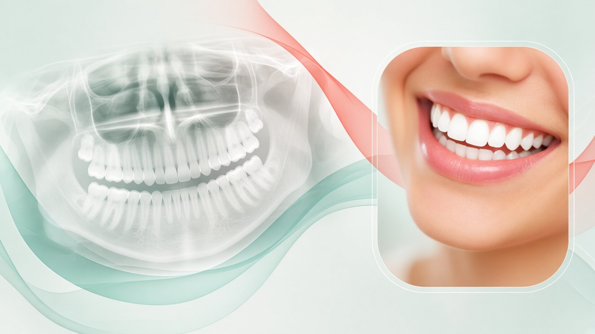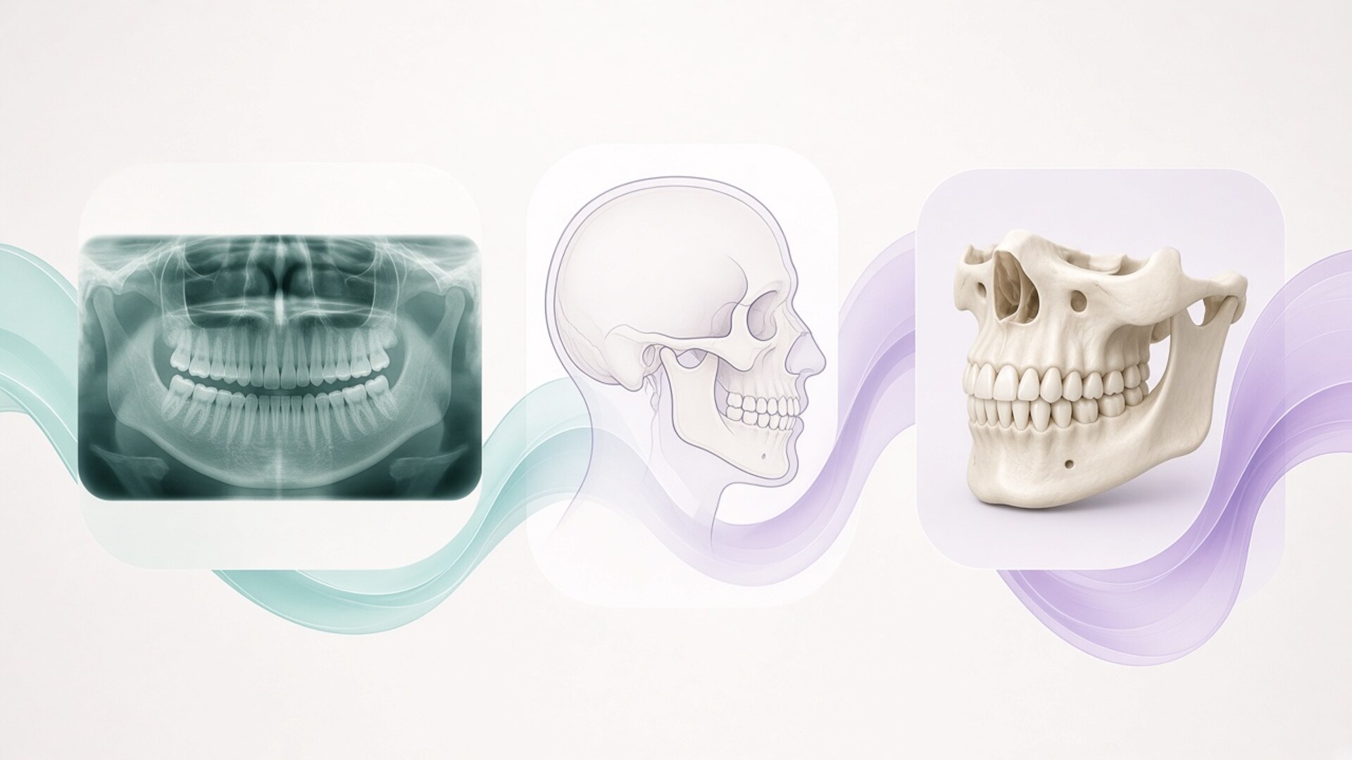
An OPG, a cephalometric X-ray and a CBCT scan answer different questions, so an orthodontist rarely picks one "best" image. I am Dr. Maksut Behruzoglu, a specialist orthodontist in Dubai, and in X-ray and CT imaging visits I explain it this way. The OPG (orthopantomogram, or panoramic X-ray) shows every tooth and both jaws in one flat picture: developing teeth, impacted teeth, bone around the roots, the rough shape of the jaw joints. The lateral ceph (cephalometric radiograph, a side view of the skull) exists for measurement: I trace angles and distances between the jaws, the skull base, the front teeth and the soft-tissue profile. CBCT (cone-beam computed tomography) gives volume: slices in any direction, bone thickness, the exact position of an impacted canine next to its neighbours. A panoramic or ceph dose usually sits in single digits to a couple of dozen microsieverts; CBCT ranges from tens to hundreds depending on the machine and field of view. That is why I order 3D for a specific clinical question. Below: what each scan shows, where each one misleads, and how they feed into a treatment plan.
Why an orthodontist may need three different images
Patients often open the consultation with one request: "Which scan should I get so everything is clear in one go?" I understand the logic. You want one test that settles the matter and saves a second trip to the imaging centre. The catch is that orthodontics works on three layers of questions, and each image sees one layer well.
The first layer is the teeth: how many there are, where they sit, whether any are missing or extra, whether there are cysts, short roots, fillings or crowns that will change the mechanics. A panoramic X-ray covers this layer well. The second layer is the skeleton: where the upper and lower jaw sit relative to the skull base and to each other, how the front teeth are angled, which direction a teenager's face is growing. That is the job of a lateral ceph with tracing. The third layer is volume: bone thickness on the lip and tongue side, the precise path of a root, the distance to a nerve, whether an impacted canine is touching the roots of the incisors. CBCT answers those.
"What is a ceph?" is the question I hear most, more than questions about the other two. Many people have had a panoramic at their general dentist, and plenty have heard of a CT scan from friends who had implants. A side view of the head looks odd by comparison: the teeth are blurry, the two halves of the jaw overlap, and the doctor draws lines all over it. For an orthodontist that image is a measuring tool, much like a tailor's tape.
I do not order all three for everyone. An adult with mild lower crowding and a balanced skeletal pattern often needs only a panoramic, photos and a scan. A teenager with a marked overbite needs a ceph, otherwise I cannot separate a tooth problem from a jaw problem. A patient with an impacted canine needs a small-field CBCT of that area, because the panoramic will not tell me whether the canine lies on the palate side or the lip side, or whether it is eating into the root of the lateral incisor. The question sets the list.
OPG: the panoramic view of both jaws
Most people recognise the panoramic by its wide, curved picture where the teeth line up in an arch, as if someone unrolled the jaw onto a flat sheet. The machine rotates around your head while the tube and detector move in sync, keeping one curved layer shaped like the dental arch in focus. Anything inside that layer comes out sharp. Anything outside it blurs, stretches or shrinks. So the OPG is excellent for an overview and poor for precise measurement. I use it as a map before planning: which teeth are present, which are still developing, what the roots and bone look like. You stand or sit for about 15 seconds, and the dose is typically 5 to 25 microsieverts. Below: what the panoramic shows, where it misleads, and when I ask for one.
What a panoramic X-ray shows
On an OPG I count teeth and check their stage of development. For a child of eight to ten this is key information: are all the adult tooth buds present, are the second premolars or lateral incisors congenitally missing, is an extra tooth sitting between the central incisors. For a teenager I look at the eruption path of the canines and at the wisdom tooth buds. For an adult I check fillings, crowns, posts, implants, root canal treatment and the bone level around the roots.
Root length and shape come through only roughly. If the upper incisor roots look clearly shorter than I would expect, I note it before any movement starts, because short roots change the forces I choose and the pace of treatment. The jaw joints appear in outline: gross asymmetry, obvious contour changes, a difference in ramus height. The maxillary sinuses and nasal septum are partly in view.
A panoramic often catches findings the patient knows nothing about: a cyst around an unerupted tooth, a lesion at a root tip, a root fragment left after an old extraction. I discuss those with the general dentist or oral surgeon before the orthodontic plan goes ahead.
Where the panoramic misleads
The curved focal layer creates distortion. If you stand slightly forward or back from the correct position, your front teeth come out wider or narrower than they really are. Tilting the chin down gives the bite plane an exaggerated "smile"; tilting it up flattens it. A small head rotation makes one side look larger than the other, and an untrained eye sees jaw asymmetry that does not exist.
Horizontal measurements on an OPG are unreliable. Vertical ones are a bit better but still carry error. So I do not use a panoramic to measure the distance to a nerve before placing a mini-screw, and I do not judge bone thickness from it. Root angulation is also distorted, particularly around the canines and premolars: roots can look as if they converge when in reality they are parallel.
Overlap is the other limit. The spine projects over the front teeth, the tongue and air spaces cast shadows, and the palate lies across the upper roots. A flat image is read with those shadows in mind, and a radiologist's report helps when the film comes from another centre.
When I ask for an OPG
Before any orthodontic treatment I need a current panoramic or an equivalent image. By current I mean taken within recent months and reflecting the teeth as they are now. For a child in the middle of losing baby teeth, a film from two years ago is already out of date.
Mid-treatment, a panoramic helps me check root parallelism before braces come off and look for signs of root shortening in patients with risk factors. I order it when there is a reason, not on a fixed calendar. After treatment a check film is sometimes useful before retainers are made or when wisdom teeth come into the discussion.
If you already have a recent CBCT of both jaws taken for another reason, I can often build a panoramic reconstruction from it and skip a separate OPG. That saves both dose and time.
What a lateral ceph is and why orthodontists trace it
"Ceph" is short for cephalometric radiograph. The tube sits roughly one and a half metres from your head, so the beam travels almost parallel and magnification stays small and predictable. That is what makes the image measurable. Orthodontists mostly take a lateral (side) view. Your head sits in a cephalostat: two ear rods rest in the ear canals, a support touches the bridge of the nose, and your teeth meet in your usual bite. The exposure takes a few seconds and the dose is usually 2 to 10 microsieverts. The film itself tells a patient very little: a skull in profile with two jaw halves overlapping. Its value appears after cephalometric analysis, when landmarks are marked, joined with lines and compared with norms. Below: how it is taken, what gets measured, and what it cannot show.
How a ceph is taken
You stand or sit in the unit, usually the same panoramic machine fitted with an extra arm for the cephalostat. The radiographer aligns your head so that the Frankfort plane (a line from the lower edge of the eye socket to the top of the ear canal) runs parallel to the floor. If the head tilts, every angle on the film shifts and the analysis loses accuracy.
I ask you to bite the way you normally bite, without pushing the lower jaw forward. Teenagers who are self-conscious about an overbite sometimes "fix" it for the photo by sliding the jaw forward. That film shows a neat jaw relationship that does not exist in real life. Lips stay relaxed too, because strained lips distort the soft-tissue profile.
A good unit places a ruler or calibration mark on the image, so the software can convert magnification into true millimetres. If you bring a ceph from another clinic without a calibration mark, I still analyse the angles but treat linear distances with more caution.
Cephalometric analysis: angles and lines
On a ceph I mark several dozen landmarks. The main ones include point S at the centre of the sella turcica, point N at the frontonasal suture, points A and B on the front contours of the upper and lower jaw near the incisor roots, the chin points, the incisor tips, and soft-tissue points on the nose, lips and chin. Joining them produces the angles orthodontists talk about.
The SNA angle shows how far forward the upper jaw sits relative to the skull base. SNB shows the same for the lower jaw. The difference, ANB, gives a first read on skeletal class: a large ANB suggests a lower jaw that lags behind, a negative ANB suggests one that sits forward. Then I look at vertical measurements, such as the steepness of the lower jaw and the ratio of front to back face height. They hint at whether the face grows "down" or "forward", which shapes the choice of mechanics.
A separate block covers the front teeth. The tilt of the upper incisors to the palatal plane and of the lower incisors to the mandibular plane tells me how much room for movement remains within the bone and where nature has already compensated for a jaw mismatch by tipping the teeth. The soft-tissue profile helps predict how the face will change if I retract or advance the incisors.
Different schools use different analyses: Steiner, Ricketts, McNamara, Downs and others. I do not stick to one. What matters more to me is the set of measurements that answers a given patient's question, and comparing the same person's films over time.
What a ceph cannot show
A lateral ceph is one projection. The left and right halves of the jaw overlap, and when the face is asymmetric the outlines double up. I cannot judge asymmetry reliably from a lateral ceph alone; that takes a frontal ceph or a CBCT.
Individual teeth read poorly. Premolars and molars from both sides merge, and decay or root condition is not visible. Bone thickness around the roots is also out of reach, because the projection sums everything along the beam's path.
The airway shows up as a dark band behind the tongue and soft palate. Some patients ask me to assess sleep apnea from it. A flat image taken standing up with the teeth together does not reflect how soft tissues behave lying down at night. A narrow airway on a ceph is a reason to talk to an ENT or sleep physician, and I do not diagnose apnea from a film.
CBCT: volume imaging and field of view
Cone-beam computed tomography works on a different principle from a panoramic or a ceph. The tube emits a cone-shaped beam, the unit makes one rotation around your head and collects hundreds of projections. Software rebuilds them into a volume that I can slice in any direction. In orthodontics this means seeing where a root lies, how much bone is on each side, and how close an impacted tooth sits to its neighbours. Dental CBCT differs from the multi-slice CT used in hospitals: the dose is usually lower and bone detail is finer. Soft tissue, ligaments and the joint disc show poorly. A small field on one area may deliver about 10–100 microsieverts, and a large full-face field on some units reaches several hundred. Below: field of view, impacted canines, and bone for mini-screws and surgery.
Field of view and voxel size
The field of view decides how much anatomy the scan captures. A small field covers a few centimetres: one canine, a group of two or three teeth, one joint. A medium field takes in one or both jaws. A large field includes the whole face, sometimes up to the skull base.
I choose the smallest field that answers the question. If the question is one impacted canine, a large field adds dose and gives no extra information about that canine. If jaw surgery is planned, a small field is useless.
The voxel, a three-dimensional pixel, sets the level of detail. A small voxel of around 0.1 mm shows fine structures better: a root crack, early resorption, a thin cortical plate. A bone overview often works at 0.2–0.3 mm, and that setting usually carries a lower dose. A referral that states the field and the aim helps the radiographer choose the protocol, so I write mine specifically: "impacted upper right canine, relation to lateral incisor root, palatal or buccal position."
Impacted canines and neighbouring roots
After wisdom teeth, upper canines are the teeth most often stuck in the bone. On a panoramic I can see that the canine has not erupted and lies at an angle. What the panoramic cannot answer are the key questions: which side it lies on, whether it touches the lateral incisor root, and whether that root is already resorbing.
CBCT answers them. The slices show whether the canine crown sits nearer the palate or the lip, how much bone covers it, and how close it comes to the adjacent roots. Studies over the past decade show that resorption of neighbouring incisor roots next to an impacted canine is noticeably more common than flat films suggest. That matters for the plan: the canine's position dictates the surgical approach, and the state of the incisor root decides which tooth to keep if the incisor's outlook is doubtful.
The same applies to other impacted teeth, extra teeth between the incisors, and lower wisdom teeth near the nerve canal. When a surgeon has to expose a tooth and I then have to pull it into the arch, a volume scan saves time and lowers the risk to its neighbours.
Bone for mini-screws, expansion and surgery
Mini-screws are temporary anchors placed in bone where there is enough bone and no roots. In most cases a clinical exam plus a periapical or panoramic image is sufficient. I add CBCT when the space between roots is tight, when I plan palatal screws to widen the upper jaw in an adult, or when a screw has to go into an unusual site.
For adult upper jaw expansion, CBCT shows palate thickness, the state of the midline suture, and how thin the bone is on the cheek side of the back teeth. If that cheek-side bone is already thin, aggressive expansion by tipping teeth outward risks gum recession. That changes the plan.
Before orthognathic surgery, many surgeons treat a large-field CBCT as standard: they use it for virtual planning, printing surgical splints and locating the nerve canal. Here the orthodontist and the surgeon work from one volume, and repeating the scan for each specialist makes no sense.
OPG, ceph and CBCT compared in one table
Below is the working cheat sheet I keep in mind during consultations. Dose figures are approximate: they depend heavily on the specific unit, settings, field of view and the patient's age. I added the periapical X-ray for comparison because it often travels alongside a panoramic. Your actual set is decided after an exam.
| Feature | Periapical X-ray | OPG (panoramic) | Lateral ceph | CBCT (3D) |
|---|---|---|---|---|
| Image type | Flat, 1–3 teeth | Flat, both jaws as a curve | Flat skull profile | Volume, slices in any plane |
| Main question | Root, decay, root tip | Overview of teeth, buds, bone | Jaw relationship, incisor tilt, profile | Root position, bone thickness, impaction |
| Good for measuring | Limited | No, horizontal distortion | Yes, angles and calibrated distances | Yes, in three planes |
| Typical dose, µSv | about 1–5 | about 5–25 | about 2–10 | 10–100 (small field) up to hundreds (large field) |
| Scan time | 1–2 seconds | 10–20 seconds | a few seconds | 5–40 seconds |
| Weak spots | Tiny area | Overlap, magnification, false asymmetry from head position | Sides overlap, teeth read poorly | Metal artefacts, poor soft-tissue detail |
| Typical orthodontic use | Checking a root, a doubtful tooth | Start of any treatment, mixed dentition | Skeletal class, growth, profile, teen follow-up | Impacted canine, tricky mini-screw sites, surgery, asymmetry |
| Can be derived from CBCT | Partly | Yes, reconstruction | Yes, synthetic ceph | Not applicable |
The table does not mean the right-hand column beats the left. CBCT gives more information at the cost of more dose and money. If a panoramic and ceph answer the question, I stay with them. If flat images leave a fork in the road that changes the plan, volume imaging earns its place.
How I match the scan to the question
International guidance on orthodontic imaging, including the joint position of the American Academy of Oral and Maxillofacial Radiology (AAOMR) with the American Association of Orthodontists (AAO), and European guidelines agree on one point: order an image when the answer will change treatment. Routine CBCT for every orthodontic patient is not part of those recommendations. I follow the same logic.
At the first consultation I look at the face, the bite, photos and the scan, and ask myself what I do not yet know and what I need to know before planning. If an adult has a balanced skeletal profile, all teeth present and a goal limited to alignment, I need a panoramic to check roots and bone. Sometimes I add a ceph if I plan to change incisor inclination noticeably and want to see where the teeth sit within the bone.
A teenager with an overbite or underbite almost always needs a ceph. It tells me whether the bite comes from jaw position or tooth position, and guides the choice between functional appliances, braces with elastics, and treatment with or without extractions. A repeat ceph a year or two later shows how the jaws responded to growth and the appliance.
I order CBCT in specific situations. An impacted canine or other tooth that flat images cannot locate. Suspected root resorption that a flat image neither confirms nor rules out. Marked facial asymmetry. Mini-screws in a tight space between roots or in the palate. Preparation for jaw surgery. Bone questions in adults with thin gums before expansion. Occasionally a joint question when the exam points to bony change; the disc itself is assessed on MRI.
Some patients ask for CBCT on their own because "it's more accurate." I explain that accuracy matters where a decision depends on it. If a CBCT will not change the appliance, the sequence or the prognosis, the extra dose and cost are hard to justify. If the patient still wants one, we talk about the smallest field that could answer at least one useful question.
The opposite happens too. A patient with an impacted canine turns down CBCT out of fear of radiation. I show what risk that shifts onto them: the surgeon exposes the tooth without a clear map, I pull the canine without knowing how it relates to the incisor root, and we find resorption later when the root has already shortened. A small-field dose in that situation is comparable to a few days of natural background radiation. The decision stays with the patient; my job is to give them the numbers.
Scans from another clinic or another country
Most of my patients in Dubai have moved from somewhere else. They bring films from London, Moscow, Istanbul, Mumbai, Almaty. I always ask for everything you have, even old images. Old films are valuable for change over time: two cephs three years apart show how a child grew, and two panoramics show whether root length has changed.
Format matters. Bring panoramic and ceph images as full-resolution digital files. A phone photo of a monitor is fine for a first impression but not for analysis, because perspective bends the angles and glare hides detail. Bring CBCT as a complete DICOM dataset, not a handful of screenshots. Screenshots show what the previous doctor chose to look at, and I need the full volume to find my own slices.
I read the radiologist's report if there is one and translate it into planning terms. Reports in English, Turkish and Russian I read myself. For other languages a translation helps, but I review the volume itself regardless of the text.
How long a scan stays useful depends on the question and on age. For an adult with a stable mouth, a year-old panoramic is often fine for a first consultation, though I may ask for a new one before treatment starts if there have been extractions, root canals or implants since. For a child who is losing baby teeth, a six-month-old film may no longer reflect where the buds are. A CBCT that clearly answered its question usually does not need repeating unless the situation has changed.
Scans taken for other specialists count too. If your implant dentist recently took a CBCT of both jaws, I ask for that file. It often covers my questions as well, and a repeat scan becomes unnecessary. That is one reason I ask patients to keep their discs and cloud links rather than throw them away.
How scans become a digital treatment plan
Images on their own neither treat nor plan. In digital treatment planning I combine them with an intraoral scan, face and smile photos, a bite record and your goals.
The scan captures the exact shape of crowns and gums but cannot see roots or bone. X-rays capture roots and bone but render the enamel surface less precisely. In software the two can be merged: roots from a CBCT align with crowns from the scan, and I can see where a root will travel when the crown moves as planned. Most patients do not need that merge. In complex cases with thin bone or impacted teeth it helps keep roots inside the bone.
The ceph sets the frame. If the analysis shows the lower incisors are already tipped well forward, I will not plan to push them further just to gain alignment, even if aligner software offers that option. If the upper jaw is narrow and the face grows vertically, the choice of mechanics reflects it. A digital tooth model without a skeletal frame can show a beautiful result that the bone will not support.
The panoramic sets tooth-level limits: short roots, large fillings, crowns, and implants that cannot move. An implant stays where it is, and the plan is built around it.
At the planning consultation I show you the images on screen. I explain what I see, where the risks are, and what options exist. Patients who see their impacted canine in 3D for the first time usually understand straight away why surgery is needed and why treatment will take longer. Timelines are easier to discuss when you are looking at the same picture I am.
I order progress images only when there is a reason. A panoramic before braces come off, to check root parallelism. A repeat ceph in teenagers, to judge growth and appliance response. A repeat CBCT is rare: to confirm the position of a canine after it has been brought into the arch, or to check a root after a demanding movement.
Imaging prices in Dubai vary by centre and by policy. If cost comes up, I give an AED range with the price-list date and remind you to confirm coverage with your insurer, because orthodontic imaging is covered in different ways across plans.
Frequently asked questions about OPG, ceph and CBCT
What is a cephalometric X-ray in plain terms?
A ceph is an X-ray of the head in profile or from the front, taken from a longer distance with the head held in a positioning device. Distance and positioning keep distortion low, so the image can be measured. The orthodontist marks reference points and calculates how the jaws, front teeth and soft-tissue profile relate. Those numbers guide the treatment approach and let us compare results later.
Can one CBCT replace an OPG and a ceph?
Technically a large-field CBCT can produce both a panoramic reconstruction and a synthetic ceph. That makes sense when CBCT is needed for its own reason, such as an impacted canine or surgical planning. Taking a large-field CBCT only to replace two flat images is usually hard to justify, because its dose exceeds a panoramic and ceph combined. The right choice depends on the questions in your case.
Why won't my orthodontist accept the panoramic from my dentist?
I accept it if the image is recent, good quality and free of major positioning errors. Sometimes a panoramic is old, taken with the head rotated, or saved at low resolution, and then I ask for a new one. For children who are losing baby teeth, even six months can make a difference. I always look at the film you bring before ordering another.
How many X-rays will I need during orthodontic treatment?
Usually a baseline set before starting, sometimes a mid-treatment check, and a panoramic before braces come off or aligners finish. Teenagers with growth-related goals may have a repeat ceph to see how the jaws respond. A repeat CBCT is rare and tied to a specific question. The exact number depends on case complexity and treatment length.
Is dental CBCT the same as a hospital CT scan?
Both produce a 3D image, but they work differently. Dental CBCT uses a cone-shaped beam and a single rotation, usually delivering a lower dose with fine bone and tooth detail. Hospital multi-slice CT shows soft tissue better and serves other purposes. For most orthodontic questions CBCT is sufficient.
Do adults need a ceph?
Often yes, if treatment will noticeably change incisor position, correct a skeletal bite, involve extractions, or combine orthodontics with surgery. The ceph shows how much room the incisors have within the bone and how the profile may change. An adult with minor crowding and a balanced profile does not always need one. I decide after the exam and photos.
Which old scans should I bring to the consultation?
Everything you have: panoramics, cephs, CBCT, periapicals, with dates. CBCT is best as a complete DICOM dataset, and panoramic or ceph images as original digital files. If there is a radiologist's report, bring it too. Old scans show change over time and sometimes spare you a repeat exposure.
Can I get scans at one centre and treatment from another doctor?
Yes, that is common in Dubai. What matters is receiving the files in good quality and full format, not only printouts or screenshots. I write a specific question on the referral so the centre picks the right protocol and field of view. If the scan misses the needed area it has to be repeated, so it is worth agreeing on the referral in advance.